DISTINGUISHED ACHIEVEMENT AWARD WINNERS
SCVP is proud to honor these pioneers and leaders of our field.
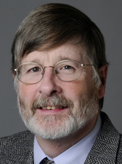
2026 - Mary Sheppard, BSc, MD, FRCPath, FRCPI
Mary Sheppard, BSc, MD, FRCPath, FRCPI
Professor Mary Sheppard’s remarkable career in cardiovascular pathology began at University College Cork, Ireland, where she earned her medical degree and an honors BSc in Pathology. She completed specialist training in pathology at Cork University Hospital, Hammersmith Hospital, and University College Hospital London, was awarded a Wellcome Fellowship for postgraduate research in 1981, and completed her MD research degree in 1984. She was trained in cardiac pathology by Professor Michael Davies at St George’s Medical School, London, and followed him in being appointed to the Chair of Cardiovascular Pathology at the same institution in 2013.
Professor Sheppard is Director of the CRY Cardiovascular Pathology Unit at City St George’s, University of London, a unique academic centre established with funding from the UK charity Cardiac Risk in the Young. She leads the unit that undertakes examination of the heart in cases of sudden cardiac death across the United Kingdom and has established a national cardiac pathology database with over 7,000 cases on file, alongside a large biobank of cardiac tissue and genetic material for research. This national referral centre provides an invaluable service to pathologists, coroners, cardiologists, and families across the UK, guaranteeing expert cardiac pathology review and a cause of death within a few weeks of referral.
Her main areas of interest are the cardiac conditions that cause sudden death in young people, which are mainly inherited, including sudden arrhythmic death syndrome, hypertrophic cardiomyopathy, dilated cardiomyopathy, and arrhythmogenic cardiomyopathy. Her work has been instrumental in connecting pathological findings at autopsy with the cardiological screening of families, creating a critical bridge between the laboratory and the clinical care of those at risk.
Professor Sheppard has published 450 peer reviewed papers, 35 book chapters, and has served on the writing panels for 12 international guidelines. She runs a recognized international training programme for pathologists, with trainees coming from the United States, Australia, Malaysia, Italy, Austria, and Denmark, and mentors cardiac pathologists and cardiovascular researchers internationally.
Her leadership in the field has been recognized by the most prestigious societies in cardiovascular medicine and pathology. She served as President of the European Association of Cardiovascular Pathology from 2016 to 2018 and has been President of the Pathology Section of the Royal Society of Medicine. She is the lead pathologist on the Executive Board of the National Pilot Study for Investigation of Sudden Cardiac Death in the United Kingdom, formed in 2020, and serves on the international grant awarding panel for the Canadian and Danish Cardiovascular Academies. She holds an honorary doctorate from the University of Copenhagen for her collaborative research work and is an honorary fellow of the Royal College of Physicians of Ireland in recognition of her research contributions. She was awarded the Stokes Medal by the Irish Cardiovascular Society and named Alumnus of the Year at University College Cork in 2018. In 2026, she received the McKenzie Medal from the British Cardiovascular Society in recognition of her contribution to British Cardiology, and she will deliver the Doniach Lecture in 2027, awarded by the Pathological Society of Great Britain and Ireland in recognition of her research contribution to UK Pathology.
Professor Sheppard’s career embodies the very spirit of the SCVP Distinguished Achievement Award. She has built something enduring, trained the next generation with exceptional generosity, and dedicated her life’s work to answering the most important question a family can ask after a sudden and devastating loss. The Society for Cardiovascular Pathology is proud to honor her with the 2026 Distinguished Achievement Award.
2025 - Cristina Basso, MD, PhD
Cristina Basso, MD, PhD
Professor Cristina Basso is one of the most distinguished cardiovascular pathologists of her generation, a physician scientist whose career has redefined the boundaries between cardiology, pathology, and translational medicine. She graduated in Medicine in 1990 from the University of Padua, an institution with a singular place in the history of medicine, home to the world’s oldest anatomical theatre and alma mater of William Harvey, the discoverer of blood circulation. It was at Padua, under the formative influence of Professor Gaetano Thiene and the legendary cardiologist Professor Sergio Dalla Volta, that Cristina first became captivated by the mystery of sudden cardiac death and the structural substrates of cardiovascular disease. She went on to complete specialist training in both cardiology and pathological anatomy, earned a PhD in Cardiological Sciences in 1996, and broadened her training through research fellowships at leading international centers including the Royal Brompton and National Heart and Lung Institute in London, the Mayo Clinic in Rochester, the Minneapolis Heart Institute Foundation, and the Cardiovascular Center at the University of Texas Medical Branch in Galveston.
She was promoted to Associate Professor at the University of Padua in 2002 and to Full Professor of Pathology in 2015, joining the Department of Cardiac, Thoracic and Vascular Sciences and Public Health. She later completed formal board certification in pathology in 2022, a credential that reflects her lifelong commitment to the discipline’s highest standards. Today she serves simultaneously as Chief of the Cardiovascular Pathology Service at Padua University Hospital, Director of the Cardiovascular Pathology Registry of the Veneto Region, Director of the Postgraduate International Master’s Course in Cardiovascular Pathology, and Vice Rector for International Relations at the University of Padua, one of the oldest and most prestigious universities in the world.
Her scientific contributions have been fundamental and far reaching. She made pioneering advances in endomyocardial biopsy and was among the first investigators worldwide to identify disease causing genes in arrhythmogenic cardiomyopathy between 2002 and 2006. Her original research brought sustained international attention to congenital anomalies of the coronary arteries, to conduction system abnormalities and interventricular pre-excitation, and to myocarditis as a cause of sudden death in the young, including a landmark series of contributions from 2000 through 2022. Her international authority in myocarditis was recognized by an invited review article in the New England Journal of Medicine in 2022, one of the most coveted platforms in all of medicine. She is first author of the landmark 2017 international guidelines paper on the investigation of sudden cardiac death, which established the role of postmortem molecular analysis, or molecular autopsy, as a standard element of modern forensic cardiac practice. She has also been a defining voice in the understanding of arrhythmic mitral valve prolapse syndrome, work she presented as the invited Douglas P. Zipes Lecturer at the 2020 Annual Scientific Sessions of the Heart Rhythm Society, the world’s largest cardiac electrophysiology organization.
In 2024, at the Congress of the European Society of Cardiology in London, Professor Basso became the first pathologist ever selected to deliver the William Harvey Lecture in Basic Science, the ESC’s most prestigious recognition for a career of transformative scientific contribution. The symmetry was profound: Harvey himself had studied at Padua, the very institution where Cristina trained and now leads. The honor was understood not only as a tribute to her personally but as a recognition of cardiovascular pathology as a discipline.
Professor Basso coordinates the North East Italy Veneto Region Network for Cardiovascular Pathology and has served as Past Chair of the ESC Working Group on Developmental Anatomy and Pathology. She served as President of the Society for Cardiovascular Pathology from 2017 to 2019 and is currently President of the Association for European Cardiovascular Pathology for the term 2023 to 2025.
Her scientific output is extraordinary by any measure. She has published hundreds of papers in the most selective journals in cardiovascular medicine and pathology, including the American Journal of Pathology, Circulation, European Heart Journal, Heart Rhythm, JACC, JAMA, The Lancet, the New England Journal of Medicine, and Virchows Archiv. She has delivered more than 300 invited lectures at national and international congresses and is the author of monographs on arrhythmogenic cardiomyopathy, cardiac tumors, and sudden death in the young and in athletes. As of May 2024, her Google Scholar h-index stands at 126 with over 98,000 citations, placing her among the most impactful researchers in the world in her field. She has received more than 14 national and international prizes over the course of her career.
At Padua she has established both an international master’s course and a PhD program in Cardiovascular Pathology, training the next generation of cardiovascular pathologists from across the globe. Her dual mastery of clinical cardiology and pathological anatomy gives her a perspective that is genuinely rare, and it is precisely this breadth that has allowed her to build bridges between the autopsy table and the cardiology clinic, between molecular genetics and morphological diagnosis, between local patient care and global scientific leadership.
Professor Cristina Basso’s career is a testament to what sustained intellectual passion, rigorous scientific discipline, and deep humanity can achieve. The Society for Cardiovascular Pathology is proud to honor her with the 2025 Distinguished Achievement Award.
2024 - E Rene Rodriguez, MD
E Rene Rodriguez, MD
Dr. E Rene Rodriguez’s journey into cardiovascular pathology began during his time in medical school, where a stint as a visiting student and later an assistant physician in Anatomic Pathology at the National Institute of Cardiology in Mexico City ignited his passion for the field. This early experience served as the foundation for his future endeavors after he obtained his medical degree from the Universidad Autónoma Metropolitana – Xochimilco in January 1982.
In August 1982, Rene embarked on a four-year fellowship at the Ultrastructure Section, Pathology Branch of the National Heart, Lung, and Blood Institute in Bethesda, MD, under the mentorship of leading experts in the field of cardiovascular pathology. Rene then transitioned into Anatomic and Clinical Pathology residency training at The George Washington University Medical Center in Washington, DC.
In 1990, Rene assumed the directorship of the Cardiovascular Pathology Unit at Rush Heart Institute in Chicago, IL. During his tenure at Rush University, he also undertook the responsibility of directing Autopsy Pathology services. In 2000, Rene took on the role of Director of Clinical Services in Cardiovascular Pathology at The Johns Hopkins Hospital in Baltimore, MD. His leadership and contributions solidified his reputation as a respected figure in cardiovascular pathology.
Since 2004, Rene has served as the Director of Cardiovascular Pathology and Director of Autopsy Pathology at The Cleveland Clinic in Cleveland, OH. In addition to his extensive professional experience, he is a Professor of Pathology at the Cleveland Clinic College of Medicine of Case Western Reserve University. His outstanding contributions to the education of cardiovascular medicine fellows was duly recognized with a teaching award in 2015.
His academic impact include more than 200 manuscripts and book chapters covering his multidimensional expertise in the field from basic science to clinical cardiology. He played a pivotal role in revising the criteria for antibody-mediated rejection in cardiac transplantation during his tenure as Chair of heart sessions at the Banff Conference in Allograft Pathology and later of the Pathology Council of the International Society for Heart and Lung Transplantation.
Throughout his career, Rene has been dedicated to advancing the field of cardiovascular pathology through his clinical expertise, leadership, and scholarly contributions. His commitment to nurturing the next generation of cardiovascular pathologists truly exemplifies the spirit of the 2024 SCVP Distinguished Achievement Award.
2023 - John Veinot, MD
John P. Veinot, MD
Dr. John Veinot is an exemplary physician, educator, investigator, and administrator whose contributions to cardiovascular pathology and to the Society for Cardiovascular Pathology span three decades of sustained excellence.
John completed his undergraduate training at Acadia University in Wolfville, Nova Scotia, before earning his medical degree from Dalhousie University in Halifax in 1988. He completed a rotating internship at Victoria Hospital in London, Ontario, followed by residency in Anatomical Pathology at Queen’s University in Kingston and a fellowship in Cardiovascular Pathology at the Mayo Clinic in Rochester, Minnesota. In 1994, he joined The Ottawa Hospital, the University of Ottawa, the University of Ottawa Heart Institute, and the Children’s Hospital of Eastern Ontario, where he would spend his entire career and build a legacy of exceptional service across all four institutions.
He rose to Full Professor in the Faculty of Medicine and held a cross-appointment to the Division of Cardiology, reflecting the breadth of his clinical expertise. His administrative record is remarkable in its scope: he served as Chair of the Department of Pathology and Laboratory Medicine at the University of Ottawa for eleven years, Head of the Departments of Pathology and Laboratory Medicine at both The Ottawa Hospital and the Children’s Hospital of Eastern Ontario, and Chief Medical and Scientific Officer of the Eastern Ontario Regional Laboratory Association from 2010 to 2021, where he championed quality and patient-centered care across the region. He also served on the Physician Advisory Panel of Accreditation Canada.
John’s passion for teaching earned him recognition at every level. He received the Faculty of Medicine Award for Excellence in Medical Education in 2001, the uOttawa Excellence in Education Prize in 2002, the Pathology and Laboratory Medicine Best Teacher Award in both 2000 and 2010, the Faculty of Medicine Educator Award for Communicator Competence in 2014, and the Physician Leadership Award from The Ottawa Hospital in 2016. He understood that the reach of good teaching extends well beyond medicine, and he took particular pride in the achievements of those he mentored throughout his career.
As a Clinical Investigator at the Ottawa Hospital Research Institute, John produced more than 250 manuscripts and multiple book chapters spanning basic science and clinicopathological correlations across the full breadth of cardiovascular disease. He co-authored a landmark book on echocardiography-pathology correlations that has served as a valued resource for cardiologists and pathologists alike.
Within the SCVP, John served on multiple committees, led the Society as its President, received the SCVP Award of Merit in 2010, and was instrumental in the publication of multiple consensus documents developed jointly with the Association for European Cardiovascular Pathology. He championed new modes of education, advanced the cause of diversity within the field, and worked tirelessly to strengthen the transatlantic bonds between the two societies, expanding the reach and influence of cardiovascular pathology on both continents.
More than any title or award, John Veinot will be remembered as someone who was always approachable, always dependable, and always willing to give his time in service of the discipline he loved. The Society for Cardiovascular Pathology is proud to honor him with the 2023 Distinguished Achievement Award.
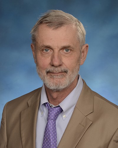
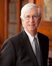
2022 - Richard Mitchell, MD, PhD
Richard N. Mitchell, MD, PhD
Dr. Rick Mitchell is an exemplary physician-scientist-educator who has made broad and impactful contributions to the field of cardiovascular pathology over many years.
Rick received his Ph.D. in Cell Biology/Immunology from The Rockefeller University in 1980 and his M.D. from Harvard Medical School in 1984. Following an internship in internal medicine at the Beth Israel Deaconess Medical Center, he completed pathology residency, cardiac pathology fellowship, and post-doctoral fellowship at the Brigham and Women’s Hospital. He joined Harvard Medical School and BWH as Assistant Professor in 1992 and ascended the academic ranks, culminating in his appointment as the Lawrence J. Henderson Professor of Pathology and Health Sciences and Technology in 2011.
Rick is a leading expert in the discipline and practice of cardiovascular pathology. He serves as a Senior Pathologist in the cardiac pathology and autopsy divisions in the Department of Pathology at BWH. He has made numerous research contributions in work conducted at the interface of immunology and vascular cell biology, including the mechanisms underlying acute and chronic rejection following heart transplantation, the role of specific cytokines and chemokine pathways in allograft vasculopathy, the source of recruited intimal smooth muscle cells in atherosclerosis, the nature of inflammatory cell populations involved in the pathogenesis of abdominal aortic aneurysm, and the immunology of cardiac valve disease. Moreover, he has been and continues to be a highly valued collaborator in studies focused on stem cell biology and the cardiovascular system, amyloidosis, gender differences in gene expression profiles, cardiac electrophysiology, and AI applications to cardiovascular pathology.
Rick also possesses a genuine, strong dedication to and aptitude for teaching and educational leadership, and serves as an important role model to medical students, pathology residents, and faculty colleagues. His teaching of residents on the autopsy and cardiac pathology services at BWH is well known and admired. He is passionate about conveying the scientific basis of medicine in both the pre-clinical and clinical segments of the undergraduate medical curriculum, and has been Director of the HST-030 Human Pathology course for over two decades (notably, this course is ranked by the HST MD students as the most effective and enjoyable course in their entire four-year curriculum). He also actively teaches or has taught in other HST courses in Cardiovascular Pathophysiology, Immunology, and Biomaterials and Tissue Engineering, as well as in the Harvard Medical School New Integrated Curriculum’s Immunology, Microbiology and Pathology course. Rick has demonstrated long-term and highly effective leadership as Associate Director of the HST Division for over two decades, during which he has directly mentored over 500 students. His teaching commitment and prowess have been recognized with seven teaching awards at Harvard Medical School and, nationally, he has received the Robbins Distinguished Educator Award from the American Society of Investigative Pathology (ASIP) and the Michele Raible Distinguished Teaching Award in Undergraduate Medical Education from the Association of Pathology Chairs.
Rick’s educational contributions have had a wide international reach. For many years he has been active in writing and editing the most widely used textbooks that educate most medical students around the world, including Robbins Basic Pathology, Robbins and Cotran Pathologic Basis of Disease, the Pocket Companion to Robbins and Cotran Pathologic Basis of Disease, and Biomaterials Science: An Introduction to Materials in Medicine. He also co-authored Robbins and Cotran Pathology Flash Cards, with an innovative design format, and co-edited a novel online book, Pathobiology of Human Disease.
Rick has given extraordinary service to the SCVP and other leading organizations in pathology. He is a past-Programming Chair and a past-President of the SCVP. He is also past-Chair of the Education Programming Committee and past-President of the American Society for Investigative Pathology, and developed and directed the annual ASIP Summer Academy, during which he taught fundamentals of acute and chronic inflammation and wound healing to graduate students, fellows, junior and senior faculty, and scientists in industry. He is one of the original organizers of the annual ASIP stand-alone meeting Pathobiology for Investigators, Students, and Academicians (PISA). Rick is also a Senior Associate Editor of the American Journal of Pathology, and Associate Editor of both Laboratory Investigation and SCVP’s official journal, Cardiovascular Pathology.
2021 - Not given
2020 - Allen P. Burke, MD
Allen P. Burke, MD
Dr. Allen Burke is one of the most prolific and influential cardiovascular pathologists of his era, a scholar whose work has shaped the field’s understanding of sudden cardiac death, coronary atherosclerosis, and cardiac tumors across more than three decades.
He earned his undergraduate degree and medical degree from the University of California, Los Angeles, graduating from the David Geffen School of Medicine in 1979. He completed his residency in Anatomic and Clinical Pathology at Letterman Army Medical Center, followed by three fellowships at the Armed Forces Institute of Pathology in Washington, DC, in genitourinary pathology, gastrointestinal pathology, and cardiovascular pathology. He went on to serve as Chair of the Department of Cardiovascular Pathology at AFIP, one of the most storied pathology institutions in the world, and later as Associate Medical Director at CVPath Institute. He is currently Professor of Pathology at the University of Maryland School of Medicine, where his research spans coronary atherosclerosis, sudden cardiac death, cardiomyopathy, cardiac tumors, and thoracic pathology.
His scientific contributions are distinguished both by their volume and their impact. Working alongside Dr. Renu Virmani and colleagues at AFIP, he produced seminal research on the mechanisms of plaque rupture and sudden coronary death, including a landmark paper published in the New England Journal of Medicine in 1997 that linked coronary risk factors to the pathological substrate of acute thrombosis. That body of work fundamentally advanced understanding of vulnerable plaque biology and remains among the most cited literature in cardiovascular pathology. More recently, he contributed to the pathological evaluation of the first genetically modified pig heart transplanted into a living human patient, a historic milestone in cardiac xenotransplantation. He served as senior editor of the 2015 WHO Classification of Tumors of the Heart and Pericardium, the definitive international reference in the field.
His scholarly output is extraordinary: more than 260 peer-reviewed publications, 6 complete books, and more than 70 book chapters, with a striking proportion carrying his name as first or last author. His work has accumulated over 21,000 citations, placing him among the most cited pathologists anywhere in the world. He has been a member of the Society for Cardiovascular Pathology and a member of its journal’s editorial board since 2002, and has contributed to the education of medical students, pathology residents, pathologists’ assistants, clinicians, and scientists throughout his career, many of whom are now active leaders in the cardiovascular pathology community.
The Society for Cardiovascular Pathology is proud to recognize the breadth, rigor, and enduring influence of Dr. Allen Burke’s contributions to the field. The Society for Cardiovascular Pathology is proud to honor Dr. Allen Burke with the 2020 Distinguished Achievement Award.
2019 - Gayle Winters
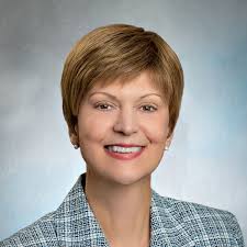
, MD
Gayle Winters, MD
Gayle Winters is a distinguished member of the global cardiovascular pathology and transplantation communities. She received her BS in Biology (with Distinction) from Purdue University, and her MD in 1979 from the Loyola Stritch School of Medicine, followed by a year of general surgery residency, residency in anatomic and clinical pathology, and fellowship in surgical pathology at Loyola University Medical Center. After joining the Loyola faculty and rising to Associate Professor of Pathology, she was recruited to the Brigham and Women’s Hospital (BWH) in 1992, where she is Senior Pathologist and Associate Professor of Pathology at Harvard Medical School.
Dr. Winters has a national and international reputation as a heart transplant pathologist and serves or has served on the Editorial Boards of Circulation, Cardiovascular Pathology, Journal of Heart and Lung Transplantation, and Advances in Anatomic Pathology. Within the Society for Cardiovascular Pathology, Dr. Winters has held important leadership positions as Cor Notes Editor, Secretary, Acting Treasurer, Vice-Present/President Elect, and President.
Among sentinel contributions to her field, Dr. Winters was a member of the working group and co-author on the original 1990 grading system for cardiac transplant biopsies, its revision in 2005, and the grading system for antibody-mediated rejection in 2011. These grading schemas are considered the foundational guidance for all of heart transplant practice to the current time. Her work demonstrated the lack of progression of untreated Grade 2 rejection, and was instrumental in prompting the revision of the grading system in 2005. Other important work within the area of cardiac transplantation includes defining perioperative ischemic injury on post-transplant endomyocardial biopsies, studying the pathologic composition and clinical significance of Quilty effect, and defining the composition of allograft coronary disease and the use of myocyte vacuolization on post-transplant endomyocardial biopsies as a predictor of allograft coronary disease.
Dr. Winters has been actively involved in resident and medical student education throughout her career. In fact, she is a major educational leader in her home institution. She was the Director of the BWH Autopsy Service for over 20 years, Director of the BWH Residency Program in Pathology from 2001-2015, and served as Director of the BWH Harvard Medical School elective clerkship in Pathology. She has taught at Harvard Medical School in the Human Pathology and Cardiovascular Pathophysiology courses. Of perhaps the greatest significance in terms of the continued expertise in cardiovascular pathology as a discipline, Dr. Winters and her colleagues at BWH have trained 12 Cardiovascular Pathology Fellows. Of these Fellows, 7 have pursued a career in cardiovascular pathology and are current members of the Society for Cardiovascular Pathology. At the national level, she has been an active member of the Program Directors for Pathology (PRODS), and served as Member-at-Large of PRODS Council, Co-Chair of the PRODS Outcomes Committee, Secretary-Elect, Secretary, Chair-Elect, and was elected Chair in 2014-15. She received the Graduate Medical Education Distinguished Teaching Award from the Association of Pathology Chairs in 2016.
In summary, Dr Winters exemplifies the professional skills and leadership qualities that the SCVP Distinguished Achievement Award was established to honor. She has been and is a recognized scholar in her field, she is widely known as an educator in cardiovascular and transplant pathology, and she has served in many leadership roles for the SCVP and other related organizations. She certainly is an expert in the field of cardiovascular pathology.
Finally, Dr Winters is an absolutely lovely human being. She is a person of great character and great courage.
2018 - Christine E. "Kricket" Seidman, MD & Jonathan Seidman, PhD
Christine E. “Kricket” Seidman, MD and Jonathan Seidman, PhD
Few partnerships in the history of cardiovascular medicine have been as scientifically consequential as that of Dr. Christine “Kricket” Seidman and Dr. Jonathan Seidman. Together, they have transformed the understanding of hereditary heart disease from the level of the gene to the bedside, building one of the most celebrated research programs in cardiovascular genetics in the world.
Dr. Christine Seidman earned her undergraduate degree in biochemistry from Harvard University and her medical degree from the George Washington University School of Medicine, completing her internship and residency in internal medicine at Johns Hopkins Hospital and her cardiology fellowship at Massachusetts General Hospital before joining Harvard Medical School. She is the Thomas W. Smith Professor of Medicine and Genetics at Harvard Medical School, an Investigator of the Howard Hughes Medical Institute, a member of the Broad Institute of MIT and Harvard, and Director of the Cardiovascular Genetics Center at Brigham and Women’s Hospital. She is a member of both the National Academy of Sciences and the National Academy of Medicine, two of the most selective honors in American science, and has authored more than 400 peer-reviewed publications.
Dr. Jonathan Seidman earned his undergraduate degree in biochemistry from Harvard University and his PhD in molecular biology from the University of Wisconsin. He completed postdoctoral training in the laboratory of Dr. Philip Leder at the National Institute of Child Health and Human Development, and has been a member of the Department of Genetics at Harvard Medical School since 1981. He is the Henrietta B. and Frederick H. Bugher Foundation Professor of Cardiovascular Genetics at Harvard Medical School and a former Howard Hughes Medical Institute Investigator, having held that distinction from 1988 to 2005. He is also a member of the National Academy of Sciences.
Together they co-direct the Seidman Laboratory, a uniquely integrated program in human molecular genetics housed within Harvard Medical School and the Cardiovascular Division of Brigham and Women’s Hospital. Their discoveries have reshaped entire fields. They identified five of the eight major genes responsible for hypertrophic cardiomyopathy, establishing it definitively as a disease of the sarcomere and demonstrating that dominant mutations in genes encoding sarcomere proteins including beta-cardiac myosin heavy chain, myosin binding protein C, troponin T and I, and alpha-tropomyosin are the root cause of the condition. They went on to show that mutations in lamin A/C, eyes-absent-4, phospholamban, myosin heavy chain, troponin T, and titin cause dilated cardiomyopathy, overturning the prevailing view that unexplained dilated cardiomyopathy was primarily attributable to unrecognized infection or toxins. Their discovery that truncating titin mutations are the single most common genetic cause of dilated cardiomyopathy, accounting for 15 to 25 percent of familial and sporadic cases, was a landmark finding that continues to shape clinical practice. They also discovered the first dominant mutations in cardiac transcription factors, specifically TBX5 and NKX2.5, that cause congenital heart defects including atrial and ventricular septal defects and tetralogy of Fallot, the most common form of cyanotic congenital heart disease. By modeling these mutations in genetically engineered mice and CRISPR-edited induced pluripotent stem cell derived cardiomyocytes, they have elucidated the molecular pathways by which each mutation drives disease and identified therapeutic targets that are now the basis of clinical trials.
The honors their work has generated are a reflection of its reach. Kricket has received the American Heart Association Basic Science Prize, the American Heart Association Research Achievement Award (the Association’s highest honor), the American Society for Clinical Investigation Award, the Glorney-Raisbeck Prize, the Schottenstein Prize, the European Society of Cardiology Gold Medal, the Vanderbilt Prize in Biomedical Science, and the Paul Dudley White Award. Together, the Seidmans have received the Pasarow Foundation Award in Cardiovascular Research, the Bristol-Myers Squibb Award for Distinguished Achievement in Cardiovascular Research, and the Institut de France Fondation Lefoulon-Delalande Grand Prix pour la Science.
Their work has carried cardiovascular pathology into a new era, one in which the molecular architecture of disease is legible, where at-risk family members can be identified before symptoms appear, and where therapy can be aimed at the very mechanisms that drive pathology. The Society for Cardiovascular Pathology is proud to honor Dr. Christine Seidman and Dr. Jonathan Seidman with the 2018 Distinguished Achievement Award.
2017 - John (Jay) T. Fallon III, MD, PhD
John (Jay) T. Fallon III, MD, PhD
Dr. John “Jay” T. Fallon III, MD, PhD has dedicated more than five decades to cardiovascular pathology, building a career of extraordinary depth across research, education, and leadership that has left a lasting mark on the field.
Jay earned his Bachelor of Arts in Biology from Providence College in 1968 and went on to receive both his MD and PhD from Albany Medical College in 1974, with his doctoral thesis already revealing his lifelong focus: it was entitled “Hemodynamics and Atherosclerosis: Endothelial Damage and Ultrasonic Arterial Lesions.” He then completed anatomic pathology residency training at Massachusetts General Hospital, Harvard Medical School, supported by an NIH Public Health Service postdoctoral fellowship, and remained at MGH for nearly twenty years, rising to Assistant and then Associate Professor of Pathology at both Harvard Medical School and the Massachusetts Institute of Technology’s Division of Health Sciences and Technology. His two decades at MGH were exceptionally productive, encompassing pioneering work in antimyosin antibody imaging of acute myocardial infarction, the immunopathology of myocarditis, cardiac tumors, and the pathophysiology of atherosclerotic plaque.
In 1994 he joined the Icahn School of Medicine at Mount Sinai in New York as Professor of Pathology, where he collaborated closely with Valentin Fuster’s group on landmark studies of macrophage infiltration in acute coronary syndromes, plaque thrombogenicity, and the mechanisms of intimal hyperplasia and restenosis after vascular interventions. He was appointed Chairman and Professor of Pathology and Medicine at New York Medical College in 2009, simultaneously serving as Director of Pathology and Laboratory Services at Westchester Medical Center in Valhalla, New York, a role he held until 2019. In 2019 he joined the Brody School of Medicine at East Carolina University as Chair and Professor of Pathology and Laboratory Medicine, continuing his commitment to teaching, research, and academic leadership.
His research contributions are remarkable in both scope and impact. Across more than 290 original publications, he has elucidated the pathogenesis of thrombogenicity in atherosclerotic plaques, illuminated the role of inflammation in plaque rupture, and probed the mechanisms of restenosis following vascular interventions. His work on the immunopathology of human myocarditis and cardiac allograft rejection helped shape the diagnostic standards the field relies on today. He has accumulated over 34,000 citations, placing him among the most cited cardiovascular pathologists in the world. He served as a member of the NIH Scientific Review Group for the Cardiovascular and Renal Study Section, as principal investigator on NIH SCOR and training grants including the NIH Thrombosis SCOR Pathology Core, as co-investigator in major vulnerable plaque studies, and as a member of the NIH Myocarditis Study Group. He has been a member of the editorial board of Cardiovascular Pathology since 1991 and received board certification in Anatomic Pathology from the American Board of Pathology in 2005.
Within the Society for Cardiovascular Pathology, Jay Fallon’s contributions are foundational. He is a Founding Member of the Society, and has served as its President, Awards Chairman, and Councilor. His commitment to the organization from its earliest days reflects the same generosity of spirit he has brought to teaching and mentorship throughout his career.
The Society for Cardiovascular Pathology is proud to honor Dr. John T. Fallon III with the 2017 Distinguished Achievement Award.
2016 - Jeffrey E. Saffitz
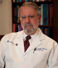
, MD, PhD
Jeffrey E. Saffitz, MD, PhD
Dr. Jeffrey E. Saffitz, MD, PhD is one of the preeminent cardiovascular pathologist scientists of his generation, whose decades of federally funded research have fundamentally advanced the understanding of sudden cardiac death and the mechanisms by which structural abnormalities in cardiac myocyte junctions give rise to lethal arrhythmias.
Jeff received his MD and PhD degrees simultaneously from Case Western Reserve University in 1978, a dual training that set the template for a career built at the interface of pathological anatomy and mechanistic science. He completed his residency in Anatomic Pathology and a Cardiovascular Research Fellowship at Washington University School of Medicine in St. Louis, then spent the 1982 to 1983 academic year as a Visiting Fellow in Cardiac Pathology at the National Heart, Lung and Blood Institute under the legendary Dr. William Roberts, one of the most formative mentors in the history of American cardiovascular pathology. He returned to Washington University as a faculty member in both the Department of Pathology and the Cardiovascular Division of the Department of Medicine, rising through the academic ranks to be named the Paul and Ellen Lacy Professor of Pathology in 1999, one of the most distinguished endowed chairs at that institution. In 2005 he was recruited to Boston, where he became Chairman of the Department of Pathology and Pathologist-in-Chief at Beth Israel Deaconess Medical Center and the Mallinckrodt Professor of Pathology at Harvard Medical School, positions he holds to this day.
His research program, continuously funded by the National Institutes of Health for several decades, has produced over 260 original contributions to the literature and established him as the world’s leading authority on the role of intercellular coupling in cardiac arrhythmogenesis. His early and sustained work on connexins, the proteins that form gap junction channels between cardiac myocytes, defined how altered expression and remodeling of connexin43 creates the substrate for lethal ventricular arrhythmias in ischemic and non-ischemic heart disease. He then extended this work into the molecular pathology of the arrhythmogenic cardiomyopathies, demonstrating that mutations in genes encoding desmosomal proteins, the structural components of cardiac myocyte mechanical junctions, disrupt the normal relationship between adhesion junctions and gap junctions and promote arrhythmogenesis. His characterization of Naxos disease, Carvajal syndrome, ARVC Type 8, and plakophilin-related cardiomyopathies established the molecular and structural basis of these conditions. In 2009 he published a landmark paper in the New England Journal of Medicine describing a novel endomyocardial biopsy test that identifies abnormally low levels of plakoglobin at cardiac myocyte junctions as a potential diagnostic marker for ARVC, a finding with direct clinical implications for the evaluation of patients at risk for sudden death.
He has authored the chapter on The Heart in the sixth and several prior editions of Rubin’s Pathology and has served on the editorial boards of ten journals spanning pathology and cardiovascular medicine. He currently serves as Associate Editor of Cardiovascular Pathology, Circulation, and the American Journal of Pathology, and as a member of the editorial board of the American Journal of Cardiology. Beyond his research and scholarship, he has been a national leader in the integration of next generation sequencing and high-throughput genomic technologies into routine clinical laboratory diagnostics, helping to bring the era of genomic pathology and personalized medicine into everyday practice.
Dr. Saffitz has served as President of the Society for Cardiovascular Pathology and in leadership roles in both the American Heart Association and the Heart Rhythm Society. He is an accomplished teacher, a generous mentor, and a model of what the cardiovascular pathologist scientist can be at the highest level.
The Society for Cardiovascular Pathology is proud to honor Dr. Jeffrey E. Saffitz with the 2016 Distinguished Achievement Award.
2015 - Michael C. Fishbein, MD
Michael C. Fishbein, MD
Dr. Michael C. Fishbein was born on May 25, 1946, in Brussels, to parents who were Holocaust survivors. With the courage of the Belgian people and the shelter of the countryside, his family endured all of World War II in hiding. In 1949 they emigrated to Chicago, where Michael’s mother had family. When Michael was twelve, his father suffered his first myocardial infarction at the age of fifty-one. From that moment, cardiovascular disease became a thread woven through the fabric of his life.
With the help of State of Illinois scholarships, Michael completed his undergraduate and medical education at the University of Illinois, where one of his teachers of congenital heart disease was the legendary Dr. Maurice Lev. He used his medical school electives purposefully and ambitiously: one took him to the Bispebjerg Hospital in Copenhagen, where he participated in an exceptionally busy autopsy service; another brought him to the Mayo Clinic, where he had the good fortune to spend time with Dr. Jack Titus. He completed his internship and residency in Anatomic and Clinical Pathology at UCLA and Harbor General Hospital in Torrance, California, where, as the only trainee with a serious interest in cardiovascular pathology, he naturally inherited the lion’s share of cardiac autopsies and built collaborative relationships with the active cardiologists on the faculty. Their enthusiasm and encouragement set the direction for everything that followed.
Recognizing that he needed deeper training, Michael was fortunate to secure an elective with Dr. William C. Roberts at the NIH, one of the most formative experiences of his professional life. There he worked alongside Dr. Victor Ferrans, Dr. Max Buja, Dr. Bernadine Healy, and Dr. Barry Maron, a constellation of future leaders in cardiovascular medicine. He also spent time with Dr. Hugh McAllister at the Armed Forces Institute of Pathology reviewing the vast congenital heart collection, and on one memorable occasion, an elephant’s heart as well.
Dr. Roberts, through his friendship with Dr. Eugene Braunwald, was instrumental in recruiting Michael to the Peter Bent Brigham Hospital in Boston, where he became the first faculty appointment of the new Chairman of Pathology, Dr. Ramzi Cotran, who famously took a considerable chance on a young man who had not yet even sat for his board exams. Michael spent three productive years at the Brigham, also consulting at Beth Israel Hospital and the West Roxbury Veterans Administration Hospital, and teaching biotechnology-oriented medical students at MIT. After the great blizzard of 1977, he and his family returned to Los Angeles, where he spent the next nineteen years at Cedars-Sinai Medical Center as head of the autopsy service and cardiovascular pathology. Practicing broad general anatomic pathology there, while demanding, gave him command of the full spectrum of human disease and a mastery of tools, including electron microscopy, histochemistry, and immunohistochemistry, that would prove invaluable to his cardiovascular research.
In 1997 he joined UCLA, where the Piansky Endowment supported his role as Distinguished Piansky Professor of Pathology at the David Geffen School of Medicine, and where the greater subspecialization of the service allowed him to focus his energies on research. He became Chief of Autopsy, Cardiovascular, and Pulmonary Pathology and holds joint appointments as Distinguished Professor in both the Departments of Pathology and Medicine. His collaborative work at UCLA extended in remarkable directions, including a productive partnership with the cardiac electrophysiology program on the role of autonomic innervation and the stellate ganglion in arrhythmogenesis, a frontier that opened new dimensions in the understanding of sudden cardiac death. His contributions to antibody-mediated rejection criteria in cardiac transplantation and to the pathology of the vulnerable atherosclerotic plaque have shaped international standards in both fields.
The scale of Michael’s scientific impact is extraordinary: more than 880 publications, an h-index of 142, and over 81,000 citations, placing him among the most cited physician scientists in the world in his discipline. Yet the numbers tell only part of the story. What defines Michael Fishbein is the warmth with which he has approached every collaboration, the generosity he has shown to trainees across five decades, and the honesty with which he has always attributed his success to good fortune, remarkable mentors, and the unwavering support of his family: his parents, who sacrificed everything so he could be educated; his wife of more than forty years, Astrid, who held everything together while he pursued his calling; and his two children, Danielle and Gregory, who have been a constant source of pride and joy.
The Society for Cardiovascular Pathology is proud to honor Dr. Michael C. Fishbein with the 2015 Distinguished Achievement Award.
2014 - Jagdish Butany
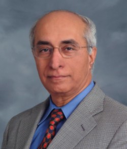
, MBBS, MS
Jagdish Butany, MBBS, MS
Professor Jagdish Butany’s career in cardiovascular pathology spans five decades and three continents, and it is a career defined above all by an extraordinary sense of purpose: a commitment to science, to teaching, to patients, and to the broader good.
Jagdish received his MBBS from Kasturba Medical College in Mysore, Manipal and Mangalore, and completed his postgraduate training at the All India Institute of Medical Sciences in New Delhi, one of India’s most prestigious medical institutions. In 1975 he moved to Toronto, where he completed his pathology training at the University of Toronto in 1979, earning his FRCPC. He then spent a transformative year as a Fellow at the National Heart, Lung and Blood Institute at the NIH in Bethesda, working under the late Dr. Victor J. Ferrans, who would become one of the most important scientific influences of his career. While at NIH he had the pleasure of working alongside Dr. William C. Roberts and a cohort of outstanding fellows, an experience that deepened his commitment to the subspecialty. On his return to Toronto, he joined the pathology service at Women’s College Hospital before moving to Toronto General Hospital as a cardiovascular pathologist, where he has remained ever since.
He is Professor in the Department of Laboratory Medicine and Pathobiology at the University of Toronto, Director of Anatomical Pathology at the University of Toronto, a role he held from 2004 to 2017, and Staff Cardiovascular Pathologist and Director of Autopsy Services at the University Health Network’s Toronto General Hospital site. He co-edited, with Dr. L. Maximilian Buja, the fourth edition of the landmark textbook Cardiovascular Pathology, which stands as one of the definitive references in the field. His more than 290 peer-reviewed publications span the breadth of cardiovascular disease, with a particular and sustained focus on prosthetic heart valves and cardiovascular devices. His rigorous analysis of prosthetic mechanical valve performance has had direct consequences for patient safety: his surveillance work on one bileaflet mechanical valve was instrumental in its recall and removal from the market in January 2000, a contribution that almost certainly saved lives.
In 2003, when SARS struck Toronto and threw its hospitals into crisis, Professor Butany stepped forward at a moment of institutional fear. At a time when many colleagues declined to participate in potentially hazardous studies, he performed autopsies on affected patients, collecting the tissue and biological evidence that helped define the pathology of the disease and support the public health response. It was an act of courage and professional responsibility that exemplified his character.
Teaching has been at the center of his identity throughout his career. He has taught medical students, pathology residents, cardiology and cardiac surgery residents and fellows, pharmacy students, nurses, physiotherapists, and occupational therapists. He built and led two highly regarded undergraduate courses in the Laboratory Medicine and Pathobiology Specialist Program at the University of Toronto, one on cardiovascular pathology and one on neoplasms, combining didactic instruction with hands-on examination of archived cardiac specimens. He has been recognized for this work with the W.H. Anderson Award for Outstanding Achievement at Toronto General Hospital in 1995, the William Aikins Award for Individual Undergraduate Medical Student Teaching from the Faculty of Medicine in 1995, the John B. Walter Teaching Award from the Department of Laboratory Medicine and Pathobiology in 2002, and the Colin R. Woolf Award for Commitment to Continuing Medical Education in 2012. He has been a visiting professor at the School of Medicine in Durban, South Africa, and has taught in CME programs across North America, Europe, and India for decades, including as a regular contributor to the programs of the Association of Indian Pathologists in North America.
His service extends well beyond his home institution. He was a founding member of the Society for Cardiovascular Pathology, which was conceived in a conference room at the University of Toronto in 1985, and served as its President from 2007 to 2009. He served as Co-Editor-in-Chief of Cardiovascular Pathology, the Society’s official journal, alongside his close colleague Professor Avrum Gotlieb, and continues to serve on its editorial board. He was President of the Canadian Association of Pathologists from 2006 to 2009, for which he received the Distinguished Service Award in 2012. He has served as Secretary-Treasurer of the World Association of Societies of Pathology and Laboratory Medicine and as a member of the Board of the World Pathology Foundation, both organizations dedicated to strengthening laboratory medicine in low-resource settings worldwide. He was involved in establishing the Royal College of Physicians and Surgeons of Canada’s Maintenance of Certification program, helping to shape the standards of professional development for Canadian pathologists for a generation.
Behind all of this is a man sustained by deep personal ties: more than four decades of love and partnership with his wife Vidya, a practising psychiatrist; the joy of watching their daughter Indrani grow; and now the pleasure of grandchildren Neveah and Riaan.
The Society for Cardiovascular Pathology is proud to honor Professor Jagdish Butany with the 2014 Distinguished Achievement Award.
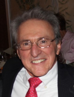
2013 - Michael A. Gimbrone Jr., MD

Michael A. Gimbrone, Jr., MD
Dr. Michael A. Gimbrone, Jr. is one of the founding figures of modern vascular biology, a physician scientist whose work over more than five decades has established the conceptual and experimental framework through which we now understand the vascular endothelium in health and disease. It is entirely fitting that he receives the Society for Cardiovascular Pathology’s Distinguished Achievement Award, following in the tradition of earlier recipients Earl Benditt, Guido Majno, Ramzi Cotran, and Russell Ross, all giants in the same intellectual lineage.
Michael received his AB in Zoology from Cornell University and his MD magna cum laude, with Honors in a Special Field, from Harvard Medical School in 1970, where he conducted research in ultrastructural anatomy and cell biology with the legendary Donald Fawcett in the Department of Anatomy. After an internship at Massachusetts General Hospital, he pursued a Research Fellowship at Children’s Hospital Medical Center in Boston with Dr. Judah Folkman, a collaboration that proved formative. Working with Folkman and another pivotal mentor, Ramzi Cotran, the young Gimbrone helped develop a rabbit cornea model that confirmed the capacity of tumors to secrete diffusible angiogenic factors, providing some of the earliest experimental evidence for what would become the field of angiogenesis. He then served as a Staff Associate at the National Cancer Institute in Bethesda before returning to Boston for residency training in Pathology at the Peter Bent Brigham Hospital, the institution that would become his permanent scientific home. He rose through the academic ranks from Instructor to Professor of Pathology at Harvard Medical School in 1985 and was later named the Ramzi S. Cotran Professor and subsequently the Elsie T. Friedman Professor of Pathology, both honoring his deep ties to the Harvard pathology tradition.
In 1976 he established the Vascular Pathophysiology Research Laboratory, and in 1998 he became the founding Director of the Center for Excellence in Vascular Biology at Brigham and Women’s Hospital and Harvard Medical School, a position he continues to hold. For approximately a decade he served as Chairman of the Department of Pathology at Brigham and Women’s Hospital, one of the most distinguished pathology departments in the world, and during much of that period he also served as the HMS Academic Dean for Partners Healthcare Systems and a member of the Council of Academic Deans at Harvard Medical School.
The scientific achievements anchored in his laboratory are of the highest order. He was among the first to establish reproducible methods for the in vitro culture of human vascular endothelial cells, a technical breakthrough so consequential that it single-handedly catalyzed the explosive growth of vascular biology as a modern research discipline. He was the first to demonstrate that endothelial cells produce prostaglandins and other bioactive mediators that govern platelet and leukocyte function, establishing the endothelium as an active participant in hemostasis and inflammation rather than a passive lining. He established the paradigm of endothelial activation by proinflammatory cytokines and discovered inducible endothelial-leukocyte adhesion molecules critical to atherogenesis, including E-selectin, now recognized as a fundamental molecule in vascular inflammation. More recently, his laboratory has led the field in understanding how biomechanical forces, particularly disturbed blood flow at arterial branch points, govern endothelial gene expression, identifying atheroprotective transcription factors including KLF2 that suppress atherogenic programs and represent potential targets for the prevention of heart attack and stroke. This arc from angiogenesis to endothelial culture to adhesion molecule discovery to mechanobiology represents one of the most coherent and productive programs in the history of vascular medicine.
His honors reflect the breadth of his influence. He has received the Basic Research Prize from the American Heart Association, a MERIT Award from the National Heart, Lung and Blood Institute, the Cardiovascular Research Lifetime Achievement Award from the Bristol-Myers Squibb Institute, the Earl Benditt Lifetime Achievement Award in Vascular Biology from NAVBO, the Warner-Lambert Parke Davis Award in Experimental Pathology from FASEB, the King Faisal International Prize in Medicine, the Okamoto Award from the Japan Vascular Disease Research Foundation, the American Society for Investigative Pathology’s Gold-Headed Cane Award in 2017, and the Frieda Robscheit-Robbins Award in 2024. He has been elected to the National Academy of Sciences, the American Academy of Arts and Sciences, and the National Academy of Medicine. He was the founding President of the North American Vascular Biology Organization.
The Society for Cardiovascular Pathology is proud to honor Dr. Michael A. Gimbrone, Jr. with the 2013 Distinguished Achievement Award.
2012 - Avrum I. Gotlieb, MD
Avrum I. Gotlieb, MD
Professor Avrum Irving Gotlieb was one of the most consequential figures in the history of cardiovascular pathology and laboratory medicine in Canada. Over more than four decades at the University of Toronto, he helped build the intellectual, educational, and institutional foundations of an entire discipline, while leaving a profound personal mark on generations of students, trainees, and colleagues who remembered him above all for his wisdom, generosity, and genuine care for the people around him.
Avrum was born in Montreal and earned his BSc in Psychology and Physiology with first class honours from McGill University in 1967, followed by his MDCM in 1971. He completed specialist training in anatomic pathology at the teaching hospitals of McGill University, receiving his Fellowship from the Royal College of Physicians and Surgeons of Canada in 1975 and certification from the American Board of Pathology in 1976. Supported by a Medical Research Council Fellowship, he then pursued postdoctoral research in the Department of Biology at the University of California, San Diego, working with the distinguished cell biologist S.J. Singer, where he developed the deep understanding of cell biology and membrane structure that would shape his research program for decades. He joined the Department of Pathology at the University of Toronto in 1978 and would remain there for the rest of his career.
He was a cardiovascular pathologist at the University Health Network and a senior scientist at the Toronto General Research Institute, whose research focused on atherosclerosis and valvular heart disease. His laboratory produced pioneering work on the role of the cytoskeleton in endothelial cell migration and repair, on the biology of valvular interstitial cells in maintaining cardiac valve structure and function, and on the mechanisms of intimal hyperplasia and its relationship to fibrofatty plaque formation. He published more than 100 peer-reviewed papers and 37 reviews and book chapters, and edited three books, including the authoritative textbook Cardiovascular Pathology, co-edited with Malcolm D. Silver of the University of Toronto and Frederick J. Schoen of Harvard Medical School.
As an institution builder, his accomplishments were remarkable. He served as the founding Chair of the Department of Laboratory Medicine and Pathobiology at the University of Toronto from 1997 to 2008, creating a new academic home for the discipline in Canada. He also served as Interim Vice-Dean for Research and International Relations, Interim Vice-Dean for Graduate Affairs, and Senior Academic Advisor to the Dean of Medicine. He was instrumental in establishing the undergraduate Specialist Program in Pathobiology, which became one of the University of Toronto’s most highly sought after life sciences programs, and helped create the MHSc in Laboratory Medicine. He established the Gotlieb Family Fund for Undergraduate Education in Pathobiology at the American Society for Investigative Pathology, providing Trainee Scholar Awards to enable undergraduates to attend the ASIP Annual Meeting each year. He authored and co-authored influential career guide booklets for ASIP, including “The Road to Becoming a Biomedical Physician Scientist in Pathology and Laboratory Medicine” and, with Dr. Tara Sander, “Journey to Success: Career Pathways for Biomedical Scientists in Pathology and Laboratory Medicine.”
His teaching was recognized with two prestigious Aikins Awards from the University of Toronto’s Faculty of Medicine, the Sustained Excellence in Graduate Teaching Award, and the Robbins Distinguished Educator Award from the American Society for Investigative Pathology in 2011. He was President of the Society for Cardiovascular Pathology from 1997 to 1999, President of the American Society for Investigative Pathology from 2001 to 2002, and President of the Canadian Society of Atherosclerosis, Thrombosis and Vascular Biology. He served as Vice President for Science Policy of the Federation of American Societies for Experimental Biology and received the Distinguished Service Award from the Association of Pathology Chairs. He was elected a Fellow of the Canadian Academy of Health Sciences in 2006. He served as Co-Editor-in-Chief of the journal Cardiovascular Pathology from 2001 to 2011, alongside his close colleague and friend Dr. Jagdish Butany, and as Associate Editor and editorial board member thereafter.
Those who knew Avrum recall not a man chasing recognition, but one focused entirely on nurturing others. He was remembered by colleagues as calm, wise, intellectually rigorous, and deeply engaged with every person he taught or mentored. His influence extended from undergraduates to department chairs, and it was felt not only in Toronto but across the international pathology community.
The Society for Cardiovascular Pathology was proud to honor Professor Avrum Irving Gotlieb with the 2012 Distinguished Achievement Award. He passed away on January 25, 2026, just days after his 80th birthday, leaving behind a legacy that will endure in the careers and lives of all those he shaped. He is deeply missed.
2011 - Gaetano Thiene, MD
Gaetano Thiene, MD
Professor Gaetano Thiene is one of the towering figures in the history of cardiovascular pathology, a physician scientist whose discoveries have saved lives on a measurable, population-wide scale and whose intellectual vision has shaped the discipline for half a century. He is Emeritus Professor of Cardiovascular Pathology at the University of Padua, the institution where he trained, built his career, and made the discoveries that made him famous.
Gaetano graduated from the Medical School of the University of Padua in 1972 and completed postgraduate training in cardiology at Padua and in pathological anatomy at the University of Trieste, earning his diplomas in 1975 and 1978 respectively. His early international training set the tone for a globally connected career: he was a Visiting Scientist at the Mayo Clinic in Rochester in 1974, at the Cardiothoracic Institute in London in 1976, and held a NATO Fellowship at the Institute of Pathology in Amsterdam in 1977. He rose through the academic ranks at Padua from Assistant Professor to Associate Professor in 1983 and to Full Professor of Pathology in 1990, a position he has held ever since. He directed the PhD Course in Cardiological Sciences and the Master in Cardiovascular Pathology from 1991, and served as Director of the Institute of Pathological Anatomy from 1998 to 2006, and as Vice-Dean of the Department of Medico-Diagnostic Sciences and Special Therapies from 2007 to 2010. He has been supported throughout his career by research grants from the European Commission, the Italian Ministry of Health, and the Italian Ministry of Education.
His most celebrated scientific achievement is his landmark 1988 paper in the New England Journal of Medicine, in which he first reported arrhythmogenic right ventricular cardiomyopathy as a major cause of sudden death in young people and athletes, a discovery that opened an entirely new field of investigation and gave generations of physicians a diagnostic category to work with. He subsequently contributed fundamentally to elucidating the genetic basis of ARVC, identifying disease-causing mutations in desmosomal genes and correlating them with the clinical and pathological features of the disease. His group in Padua has been among the world’s most productive in defining the molecular pathology of the cardiomyopathies, and Thiene himself has contributed broadly to valvular heart disease and the pathology of tissue valve substitutes, including the landmark observation that calcification in human bioprostheses is initiated in cells, as well as to myocarditis, congenital heart disease, coronary artery disease, conduction system pathology, cardiac tumors, endomyocardial biopsy, and cardiac transplantation.
Perhaps the most consequential of all his contributions, however, is the role he played as a principal architect of the Italian national preparticipation screening program for young competitive athletes. Working with colleagues Domenico Corrado and Cristina Basso, he helped design and implement the world’s most successful such program, which requires cardiovascular evaluation including ECG of all young Italians wishing to participate in competitive sports. The results have been nothing short of extraordinary: over the study period in the Veneto region, sudden cardiac death in athletes fell by approximately 90 percent, directly attributable to the identification and disqualification of young people with underlying cardiomyopathies. This program is now a model cited around the world in debates about athlete screening policy, and its scientific documentation across multiple landmark publications stands as one of the most compelling demonstrations of preventive medicine’s power in cardiovascular disease.
Professor Thiene has published nearly 900 reports of original investigations and reviews in the leading journals of pathology and cardiovascular medicine, accumulating over 67,000 citations. He has served as Associate Editor of Cardiovascular Pathology and the Journal of Heart Valve Disease, and on the editorial boards of many other prestigious journals. He was President of the Society for Cardiovascular Pathology from 2003 to 2005 and President of the Association for European Cardiovascular Pathology from 2004 to 2006, and has been a tireless champion of the subspecialty of cardiovascular pathology internationally, mentoring young European cardiovascular pathologists and leading the definition of educational and training standards for the field.
His honors have come from every major society in cardiovascular medicine and pathology. They include the Andreas Grüntzig Lecture and Award from the Swiss Society of Cardiology in 2008, the Excellence in ARVC Award from Johns Hopkins University in 2010, the Paul Dudley White International Lecture and Award from the American Heart Association in 2013, the Leonardo da Vinci Lecture and Award from the Italian Society of Cardiology in 2013, the René Laennec Lecture and Award from the European Society of Cardiology in 2014, the Commencement Lecture at the 250th Anniversary of the University of Pennsylvania Medical School in 2014, the Ruysch Lecture and Award from the Academic Medical Center in Amsterdam in 2015, and the Lady Estelle Wolfson Lecture from the Royal College of Physicians in London in 2017. He is an Honorary Fellow of the Royal College of Physicians in London, a Foreign Member of the Serbian Academy of Sciences and Arts, a Corresponding Member of the Galileian Academy in Padua, and Vice-President of the Olympic Academy in Vicenza.
The Society for Cardiovascular Pathology is proud to honor Professor Gaetano Thiene with the 2011 Distinguished Achievement Award.
2010 - Bruce McManus, MD, PhD
Bruce McManus, MD, PhD
Dr. Bruce McManus is one of the most decorated and broadly accomplished cardiovascular pathologists that Canada has produced, a physician scientist whose career has spanned basic research, clinical pathology, institution building, mentorship, and public policy in equal measure.
Bruce earned his BA in Biology and Physical Education and his MD from the University of Saskatchewan, his MSc in Applied Physiology from Pennsylvania State University, and his PhD in Exercise Physiology and Biochemistry from the University of Toledo. He pursued postdoctoral fellowships in Environmental Physiology at the University of California, Santa Barbara, and in Cardiovascular and Pulmonary Pathology at the National Heart, Lung, and Blood Institute in Bethesda, Maryland, before completing residency training in Internal Medicine and Pathology at the Peter Bent Brigham Hospital, Harvard University. He then spent eleven years on the faculty of the University of Nebraska Medical Center, including a sabbatical at the Max Planck Institute for Biochemistry in Martinsried, Germany, that led to the co-recipient Max Planck Research Award in 1991, one of the most prestigious international prizes in the biomedical sciences. In 1993 he was recruited to the University of British Columbia as Head of the Department of Pathology and Laboratory Medicine, a position he held until 2000, and where he would remain for the rest of his career, eventually becoming Professor Emeritus.
At UBC he built one of Canada’s most productive cardiovascular research ecosystems, serving as Director of the James Hogg Research Centre at St. Paul’s Hospital, Co-Director of the Institute for Heart and Lung Health, and founding CEO of the Centre of Excellence for Prevention of Organ Failure, known as the PROOF Centre, a nationally funded translational research organization dedicated to developing biosignature-based tools for the early detection and prevention of organ failure. He co-founded the Personalized Medicine Initiative of British Columbia and participated in multiple life sciences and technology start-ups, carrying his scientific insights into commercial and policy environments. His work advancing trans-omic biosignatures has been among the most innovative in the field.
His core scientific passion has been understanding the mechanisms, consequences, detection, and prevention of injury and aberrant repair in inflammatory diseases of the heart and blood vessels, with a particular and sustained focus on the diagnosis and management of acute viral myocarditis. Over the course of his career he has authored more than 400 peer-reviewed publications, contributed to more than 60 book chapters, and edited four books, with an h-index of 85 reflecting work that has touched virtually every corner of cardiovascular pathology.
The recognition he has received is among the most extensive of any Canadian cardiovascular pathologist. Beyond the Max Planck Research Award, he was elected a Fellow of the Royal Society of Canada in 2002, received the UBC Killam Research Prize as Senior Scientist in 2003, was elected an inaugural Fellow of the Canadian Academy of Health Sciences in 2005, and received the Research Achievement Award of the Canadian Cardiovascular Society that same year. In 2006 he received the BC Innovation Council’s Lieutenant Governor’s Technology Innovation Award. He was honored with the UBC Distinguished Medical Lecturer Award in 2007 and the David F. Hardwick Lifetime Achievement Award in 2008. In 2009 he received the Scientific Excellence Award from the Canadian Society for Atherosclerosis, Thrombosis and Vascular Biology. In 2015 he received the UBC Faculty of Medicine Distinguished Achievement Award for Senior Faculty, the Canadian Blood Services Lifetime Achievement Award, the Howard Morgan Award from the International Academy of Cardiovascular Sciences, and the Jacob Biely Faculty Research Prize from UBC. In 2017 the UBC Faculty of Medicine honored him with the William and Marilyn Weber Lifetime Achievement Award. In 2018 he was appointed a Member of the Order of Canada, the nation’s highest civilian honor, and received the Margolese National Heart Disorders Prize.
He served as President of the Society for Cardiovascular Pathology and as Councilor for both the International Society for Heart Research and the American Society for Investigative Pathology. He has been universally celebrated as an extraordinary mentor, described by those he trained as a scientist who invested in people with the same energy he brought to ideas.
The Society for Cardiovascular Pathology is proud to honor Dr. Bruce McManus with the 2010 Distinguished Achievement Award.
2009 - L. Maximilian Buja, MD
L. Maximilian Buja, MD
Dr. L. Maximilian Buja is one of the most enduring and multidimensional figures in the history of American cardiovascular pathology, a physician scientist and academic leader whose contributions to research, education, and institutional life span more than five decades and have left a permanent mark on the field.
Max earned his medical degree with honors from Tulane University School of Medicine in 1967, along with a Master of Science in Anatomy, and launched his career with an internship at Charity Hospital in New Orleans. He then joined the National Heart and Lung Institute in Bethesda as a Staff Associate, completed pathology residency at the National Cancer Institute, and rose to Senior Investigator in Cardiovascular Pathology at the National Heart and Lung Institute, where he was shaped by immersion in some of the most productive cardiovascular research of his era. In 1974 he joined the faculty of the University of Texas Southwestern Medical Center in Dallas, where he spent fifteen highly productive years before being recruited to McGovern Medical School at The University of Texas Health Science Center at Houston in 1989.
At UTHealth, the breadth of his administrative service has been extraordinary. He served as Chair of the Department of Pathology and Laboratory Medicine from 1989 to 1996, holding the Distinguished Chair in Pathology and Laboratory Medicine, and was appointed Dean of McGovern Medical School from 1996 to 2003, during which time he was named the H. Wayne Hightower Distinguished Professor in the Medical Sciences in 2000. He subsequently served as Executive Vice President for Academic Affairs at UTHealth and as Executive Director of the Houston Academy of Medicine and Texas Medical Center Library. Throughout all of these roles he maintained an active research program, clinical practice, and teaching presence. He has served as Chief of Cardiovascular Pathology Research at the Texas Heart Institute and as Consultant in Cardiovascular Pathology to the Harris County Institute of Forensic Sciences, and directs a cardiovascular pathology fellowship program approved by the Texas Medical Board. He served as Editor-in-Chief of Cardiovascular Pathology, the official journal of the Society for Cardiovascular Pathology, from 2012, and was President of the Society from 1987 to 1988, receiving its Award of Merit in 1998.
His research has centered on the pathobiology of cardiovascular diseases, with particular focus on myocardial ischemic and hypoxic injury, atherosclerosis, and cardiomyopathies, combining experimental cell biology with clinicopathological correlation across decades of NIH-funded investigation. His current work examines cardiac remodeling in advanced heart failure through the study of explanted hearts and left ventricular core samples obtained at the time of ventricular assist device placement, extending his contribution to the frontier of heart failure pathology. He has authored more than 500 peer-reviewed publications, three atlases of pathology, and a comprehensive textbook of cardiovascular pathology, accumulating over 19,000 citations with an h-index of 75.
His dedication to education has been as consequential as his science. He was appointed the first University of Texas Chancellor’s Health Fellow in Education in 2004, co-founded and served as founding President of the UT System Kenneth I. Shine MD Academy of Health Science Education in 2005, was named a Distinguished Teaching Professor in the University of Texas System in 2007, and founded the Shine Academy to promote excellence in health sciences education across the state. He received the UT Health President’s Scholar Award for Excellence in Teaching in 2014 and the Outstanding Alumnus Award from the Tulane Medical Alumni Association in 2008.
Among his many other honors, he was elected Fellow of the American Association for the Advancement of Science in 1997, received the Harlan J. Spjut Award from the Houston Society of Clinical Pathologists in 1997, the George T. Caldwell MD Distinguished Service Award from the Texas Society of Pathologists in 2005, and has been recognized through visiting professorships and lectureships at institutions across the United States and internationally.
The Society for Cardiovascular Pathology is proud to honor Dr. L. Maximilian Buja with the 2009 Distinguished Achievement Award.
2008 - William D. Edwards, MD
William D. Edwards, MD
Dr. William D. Edwards was born in 1948 and completed both his undergraduate and medical education at the University of Kansas. He pursued residency training in Anatomic Pathology at Kansas University Medical Center, during which he undertook a year of subspecialty training in cardiovascular pathology from 1976 to 1977 under the legendary Dr. Jesse E. Edwards at United Hospital in St. Paul, Minnesota. Studying with Jesse Edwards was itself a formative privilege: William’s mentor was among the greatest cardiovascular pathologists in American history, the architect of a registry of over 22,000 donated hearts and the co-founder of the discipline as a modern subspecialty, who had himself trained with the first open-heart surgery team at Mayo Clinic a generation earlier. William took that tradition forward, earning his Board Certification in Anatomic Pathology in 1978 and joining the staff of the Mayo Clinic in Rochester that same year, where he would spend his entire career.
At Mayo, he led the Cardiovascular Pathology Working Group and, in 1981, established the Mayo Clinic Cardiovascular Pathology Fellowship, the first ACGME-accredited training program of its kind in the United States. In doing so, he created the institutional template that the field has followed ever since, ensuring that cardiovascular pathology would be recognized, taught, and practiced at the highest level of formal medical training. He built the fellowship into one of the most respected in the world and trained nearly two dozen cardiovascular pathologists whose own careers now span the globe and continue his legacy.
His scientific contributions have been defined by exceptional breadth and rigor. He was a pioneer in the systematic pathologic evaluation of cardiovascular surgical specimens, developing standards and methods that transformed the field’s capacity to correlate operative findings with clinical outcomes and guide future surgical planning. His 1988 paper on photographic documentation of cardiovascular pathology specimens became a reference standard for the field, codifying techniques of dissection, fixation, and imaging that educators still rely on today. He worked productively at the interface of cardiovascular pathology and clinical echocardiography, a collaboration that earned him the Acoustic Visionary Award from the American Society for Echocardiography in recognition of how deeply pathological insight had enriched echocardiographic interpretation at Mayo. His publications, appearing in the New England Journal of Medicine, Circulation, the Journal of the American College of Cardiology, and dozens of other leading journals, number more than 500 peer-reviewed articles, alongside nearly as many monographs and book chapters. They have been cited over 30,000 times, and his h-index of 99 places him among the most impactful cardiovascular pathologists in the world.
As a teacher, he was beloved at every level. He received the Faculty Service Award from Mayo Medical School in 1986, the Outstanding Teacher Award from the Mayo Clinic Cardiology Fellows in 1993, 1995, and 1996, the Academic Faculty Award from the Diagnostic Echocardiography Program in 2001, and the Teacher of the Year Award from Mayo Medical School in 2002. He also received the Distinguished Educator Award from the Mayo Clinic. A sought-after lecturer at both cardiology and pathology conferences around the world, he brought the same meticulous care to teaching that he brought to dissection and documentation.
The Society for Cardiovascular Pathology is proud to honor Dr. William D. Edwards with the 2008 Distinguished Achievement Award.
2007 - Renu Virmani, MD
Renu Virmani, MD
Dr. Renu Virmani’s path to becoming one of the most consequential cardiovascular pathologists in the world began with a chance encounter. She had earned her MD from Lady Hardinge Medical College at Delhi University in New Delhi in 1973 and arrived in the United States in 1974 after marrying, having already completed her postgraduate training in pathology in India. Two months of searching for work yielded nothing until a conversation with her parents-in-law led to an introduction to Dr. Sylvan Weinberg, the influential cardiologist and past president of the American College of Cardiology, who in turn introduced her to Dr. William C. Roberts at the National Institutes of Health. That meeting changed everything. She found her intellectual home in the cardiovascular pathology laboratory at the NHLBI, completed her residency at George Washington University Medical Center in 1977 and a fellowship at the National Heart, Lung, and Blood Institute in 1979, then returned to NHLBI as a staff fellow before joining the Armed Forces Institute of Pathology as a staff pathologist.
She left AFIP briefly for Vanderbilt University, where she served as Associate Professor and Director of the Autopsy Service and directed autopsy services at the Veterans Administration Medical Center in Nashville. She also served in the U.S. Army Reserves, based at Walter Reed Army Medical Center, until 1989. But it was her return to AFIP in 1984 as Chair of the Department of Cardiovascular Pathology, a position she held for twenty years, that defined the first great chapter of her scientific career. There, working with one of the most extraordinary collections of human cardiovascular tissue in the world, she and her colleagues produced a body of research that reshaped the field.
Her greatest scientific contributions center on the pathobiology of coronary thrombosis and atherosclerotic plaque. She was the first to describe and characterize the thin-cap fibroatheroma as the morphological substrate of the vulnerable plaque, demonstrating that it is the precursor lesion responsible for plaque rupture and the majority of cases of acute myocardial infarction. She and her team were also the first to establish that coronary thrombosis does not arise exclusively from plaque rupture, but also from plaque erosion, particularly in younger women, and from calcified nodules in older patients, a conceptual advance that fundamentally broadened the understanding of how and why hearts stop. The classification of atherosclerotic lesions she helped develop with colleagues at AFIP remains among the most widely cited frameworks in the field. She was a pioneering voice in warning about the dangers of brachytherapy before they became apparent to others, and she was among the first to predict that drug-eluting stents would be associated with late stent thrombosis, poor endothelialization, and in-stent neoatherosclerosis, concerns that proved correct and led to important improvements in device design and patient management.
In 2005, Dr. Virmani founded CVPath Institute in Gaithersburg, Maryland, an independent nonprofit organization that has become one of the world’s foremost cardiovascular pathology research centers. The institute has advanced understanding of vulnerable plaque biology, stent healing, transcatheter valve pathology, and the pathophysiology of chronic total occlusion, while training fellows from around the world and conducting philanthropic work alongside medical examiners. She serves as President Emeritus of the institute, and continues as Clinical Professor of Pathology at Georgetown University, George Washington University, the University of Maryland, and Vanderbilt University.
She has delivered more than 800 presentations at scientific meetings worldwide, authored or co-authored more than 600 peer-reviewed publications, edited 7 books, and written more than 100 book chapters. Her work spans Circulation, the Journal of the American College of Cardiology, and dozens of other leading journals on whose editorial boards she has served. Among her many honors, she received the Joint Service Commendation Medal for Outstanding Meritorious Service as Chief of Cardiovascular Pathology at AFIP, the Gold Medal for Best Scientific Exhibit at the American Society of Clinical Pathologists in 1981, the Edward Rhodes Stitt Lecture Award in 1988, the TCT Career Achievement Award from the Cardiovascular Research Foundation in 2012, and the Gold Medal of the European Society of Cardiology in 2021, making her the first pathologist ever to receive that distinction. She is a Fellow of the American College of Cardiology and a member of the American Heart Association and the US and Canadian Academy of Pathology.
Bill Roberts, her first mentor, once described her simply: “She’s not a patsy.” That independence of mind, the willingness to say what the data showed even when the field did not want to hear it, has been the defining quality of her career.
The Society for Cardiovascular Pathology is proud to honor Dr. Renu Virmani with the 2007 Distinguished Achievement Award.
2006 - Frederick J. Schoen, MD, PhD
Frederick J. Schoen, MD, PhD
Dr. Frederick J. Schoen occupies a singular position in the history of cardiovascular pathology, standing at the junction of medicine, engineering, and biology in a way that few physician scientists ever have. His career has been defined by a conviction that understanding the failure of cardiovascular devices requires the same rigor and imagination as understanding the failure of natural tissues, and by decades of work that have made him the world’s foremost authority on the pathology of heart valve substitutes and cardiovascular prostheses.
Fred’s training reflects the intellectual range that would define his career. He earned a BSE in Materials and Metallurgical Engineering from the University of Michigan, a PhD in Materials Science from Cornell University, and an MD from the University of Miami School of Medicine, completing his residency in Anatomic Pathology and a fellowship in Thoracic and Cardiovascular Pathology at the University of Florida. He joined Brigham and Women’s Hospital and Harvard Medical School in 1980 as Director of Cardiovascular Pathology, where he has remained ever since, serving over the years as Vice Chairman of Pathology, Acting Chairman, and ultimately Executive Vice Chairman and Director of Cardiac Pathology. He is Professor of Pathology and Health Sciences and Technology at Harvard Medical School, a faculty member in the Harvard-MIT Division of Health Sciences and Technology, and a John F. and Virginia B. Taplin Faculty Fellow in Health Sciences and Technology. He has also served as BWH liaison to the Center for Integration of Medicine and Innovative Technology, reflecting his lifelong commitment to translational research at the interface of pathology and biomedical engineering.
His research has centered on the pathology of host-biomaterial interactions, structure-function-pathology correlations in heart valve substitutes, calcification of bioprosthetic tissues, cardiovascular tissue engineering, and heart transplantation. More than half of his more than 470 peer-reviewed publications, book chapters, and reviews are focused on heart valves, a sustained body of work without parallel in the field. He is the sole author of Interventional and Surgical Cardiovascular Pathology: Clinical Correlations and Basic Principles, published in 1989, co-edited the landmark textbook Biomaterials Science: An Introduction to Materials in Medicine with Ratner, Hoffman, and Lemons through three editions, and co-edited Silver’s Cardiovascular Pathology. Together these volumes have educated generations of pathologists, cardiologists, and biomedical engineers. He co-directed, with Dr. Michael Gimbrone, the major continuing education course in Cardiovascular Pathology at the US-Canadian Academy of Pathology for many years.
His teaching has been recognized repeatedly, including with the Irving M. London Teaching Award from the Harvard-MIT Division of Health Sciences and Technology and the 2015 Senior Faculty Mentor Award from the Brigham and Women’s Hospital Center for Faculty Development and Diversity. He is a Founding Fellow of the American Institute of Medical and Biological Engineering and has served as Vice-Chair of the Massachusetts Medicolegal Investigations Commission. He has been Past President of the Society for Biomaterials, the Society for Cardiovascular Pathology, and the International Society for Applied Cardiovascular Biology.
Among his many honors, he received the Society for Biomaterials Clemson Award for Applied Biomaterials Research in 1990, the SFB Founders Award for Lifetime Achievement in Biomaterials in 1999, the Distinguished Alumni Award from Cornell University’s Department of Materials Science and Engineering in 2011, and the Society for Biomaterials Technology Innovation and Development Award in 2015.
The Society for Cardiovascular Pathology is proud to honor Dr. Frederick J. Schoen with the 2006 Distinguished Achievement Award.
2005 - Thomas N. James, MD
Thomas N. James, MD
Dr. Thomas N. James was one of the most complete physician scientists of twentieth century American cardiology: a clinician, investigator, teacher, and institution builder of the first rank, whose discoveries in the anatomy of the coronary arteries and the cardiac conduction system shaped modern understanding of how the heart beats, and why it sometimes stops.
Tom was born on October 24, 1925, in Amory, Mississippi. A bout with malaria at the age of twelve, when a nurse showed him his own blood under a microscope, first ignited his passion for medical science. He won a Westinghouse scholarship to Tulane University in New Orleans, earning his BS in 1946 and his MD in 1949. He completed his internship and residency at Henry Ford Hospital in Detroit, then served as a Captain in the United States Army Medical Corps stationed in Osaka, Japan. Returning stateside, he joined the Ochsner Clinic in New Orleans in 1955, before Henry Ford Hospital recruited him back to head their cardiology department in 1959. In 1968 he joined the University of Alabama at Birmingham, where he would spend nearly two decades and serve as Chairman of the Department of Medicine from 1973. In 1987 he was chosen as President of the University of Texas Medical Branch in Galveston, the first and for sixty years the only medical school in Texas, and the institution from which he retired in 2004.
His seminal 1961 textbook, Anatomy of the Coronary Arteries, established his international reputation and laid the scientific foundation for treatments such as coronary artery bypass surgery and stenting. His focus then shifted to the electrical conduction system, where his meticulous postmortem anatomic studies, conducted on over 1,100 human hearts and several hundred animal hearts and documented through more than 500,000 photomicrographs, traced previously unknown internodal pathways and revealed bypass tracts that alter the behavior of the atrioventricular node. Two of these electrical pathways bear his name. The discoveries earned him global prominence and contributed directly to refinements in cardiac pacemaker design. His later research on long-QT syndrome, sudden cardiac death, and crib death, the subject of a monograph published in 2007, extended his contributions across cardiology, sports medicine, and primary care, and has continued to stimulate genetic and pathological research around the world.
Throughout his career he accumulated more than 472 publications and was recognized by the Institute of Scientific Information as one of the 1,000 most-cited scientists in the world for the period 1965 to 1978, with one of his publications in the American Heart Journal formally designated a Citation Classic. He received more than 200 awards and invited lectureships on every continent, including the inaugural Distinguished Scientist Award of the American College of Cardiology in 1982, the Theodore and Susan Cummings Humanitarian Award from the ACC in 1975, and the American Heart Association’s James B. Herrick Award in 1999. He was elected a Master of the American College of Physicians and an Honorary Member of the Chilean Society of Cardiology and Corresponding Member of the British Cardiac Society, among many international distinctions.
His leadership of the field was extraordinary in scope. He served as President of the American Heart Association, President of the International Society and Federation of Cardiology, and in 1986 presided over the Tenth World Congress of Cardiology in Washington DC, only the second American ever so honored. From 1988 to 1998 he served as Director of the Cardiovascular Center of the World Health Organization, making frequent visits to Geneva and working to shape global cardiovascular policy.
Dr. Thomas N. James passed away at his home in Birmingham, Alabama on September 11, 2010, at the age of 84. He left behind two generations of cardiologists who trained under his example, a collection of hearts and slides that remains an irreplaceable scientific resource, and a body of work that changed the way the world understands the human heart.
The Society for Cardiovascular Pathology was proud to honor Dr. Thomas N. James with the 2005 Distinguished Achievement Award.
2004 - Anton E. Becker, MD

Anton E. Becker, MD
Professor Anton E. Becker devoted 35 years of his career to building one of Europe’s most respected cardiovascular pathology programs at the Academic Medical Center of the University of Amsterdam, where he served as Professor and Head of the Department of Cardiovascular Pathology. His tenure in Amsterdam transformed the institution into an internationally recognized center for cardiovascular pathology research and training, attracting collaborators and trainees from across the world.
Professor Becker is perhaps best known for his foundational contributions to the understanding of the cardiac conduction system. His collaborations with Robert Anderson, Giel Janse, and Hein Wellens at the University of Amsterdam provided the basis for important subsequent contributions to the understanding of cardiac arrhythmias and their anatomic and functional relationships. Together with Anderson, he co-authored the landmark monograph “The Conduction System of the Heart: Structure, Function and Clinical Implications,” which became a foundational reference for pathologists, electrophysiologists, and cardiac surgeons worldwide and remains highly cited to this day.
His meticulous anatomical work on the atrioventricular node and its relationship to Koch’s triangle was particularly impactful in the era of catheter ablation. By carefully reconstructing the compact AV node in serial sections from autopsy hearts and characterizing the variation in its inferior extensions, Becker provided electrophysiologists with the anatomical roadmap needed to safely and effectively perform ablation procedures for supraventricular arrhythmias. This work exemplifies the direct and practical clinical value of rigorous cardiovascular pathology research.
Beyond the conduction system, Professor Becker made important contributions to the understanding of coronary artery disease and atherosclerosis. His work addressed the role of inflammation in coronary artery disease and helped lay the scientific groundwork for subsequent plaque vulnerability research.
Professor Becker was also a passionate advocate for the formalization of cardiovascular pathology training in Europe. His 1994 publication in Virchows Archiv titled “A European School for Cardiovascular Pathology” was a visionary call to action that predated the formal training structures later developed by the Association for European Cardiovascular Pathology, of which he was a driving force. He understood early on that the future of the specialty depended on creating structured pathways to train the next generation of cardiovascular pathologists across the continent.
After 35 years of extraordinary service to the field, Professor Becker retired from the Academic Medical Center in Amsterdam in 2004, marked by a farewell symposium organized in his honor by colleagues and former trainees from around the world, a testament to the deep respect and affection he inspired throughout his career.
The Society for Cardiovascular Pathology was proud to honor Professor Anton E. Becker with the 2004 Distinguished Achievement Award, recognizing a career defined by scientific rigor, collaborative generosity, and an enduring commitment to advancing cardiovascular pathology as both a discipline and a profession.
2003 - Keith A. Reimer, MD, PhD
Keith A. Reimer, MD, PhD
Dr. Keith A. Reimer was born in Beatrice, Nebraska in 1945, and from an early age showed the intellectual precision and curiosity that would define one of the most consequential research careers in the history of cardiovascular pathology. He earned both his MD and PhD in Experimental Pathology from Northwestern University, where he formed the scientific partnership that would shape his life’s work, teaming up with Dr. Robert Jennings, whose rigorous approach to experimental myocardial ischemia would prove transformative. When Jennings moved to Duke University in 1975, Reimer accompanied him, and the two established a laboratory that would become one of the most productive in cardiovascular biology.
Their first great contribution was the wavefront theory of myocardial necrosis. Using a canine model of coronary artery occlusion, they demonstrated that myocardial infarction does not occur uniformly or instantaneously, but progresses as a wavefront from the subendocardium toward the subepicardium, beginning as early as 20 minutes after occlusion and reaching more than 70 percent transmural necrosis by six hours. They further showed that restoring coronary flow within two hours could salvage substantial myocardium and preserve cardiac function. The implications were profound. As Duke cardiologist Dr. Dan Mark later observed, this concept “transformed cardiology from a rather passive field of observational medicine to an aggressive interventional field and has improved the lives of millions of people.” The wavefront theory provided the scientific rationale for thrombolytic therapy and, ultimately, for the modern era of percutaneous coronary intervention.
Keith’s second towering contribution came through one of his postdoctoral fellows, Charles Murry, whose 1986 paper in Circulation, produced under Reimer’s mentorship, described ischemic preconditioning: the counterintuitive discovery that brief, repetitive episodes of ischemia and reperfusion protect the heart against a subsequent sustained ischemic insult by delaying lethal cell death for an extended period. The phenomenon overturned prevailing assumptions about the vulnerability of reversibly injured myocardium and launched a field of its own. By the late 1990s, approximately 200 papers on preconditioning were being published annually, and Keith took enormous pride in Murry’s achievement and the research it had inspired. He also developed the AMPIM study, a framework for rigorously evaluating cardioprotective therapies using animal models of ischemia, which set methodological standards for the field.
At Duke, Reimer served as Chief of Cardiovascular Pathology, succeeding his mentor Donald Hackel, and was known as an outstanding teacher and a demanding but generous mentor to a generation of postdoctoral fellows and medical students who have since built distinguished careers of their own. He held leadership positions in the American Section of the International Society for Heart Research, serving as Secretary and President, and was active in shaping the direction of international cardiovascular research.
Those who knew Keith remembered him above all for the sharpness and speed of his intellect. As a colleague wrote in his obituary, his capacity to size up a problem or a concept and assess its value in an instant was his greatest talent. Beyond science, he was a devoted husband to his wife Susan and father to their son Kerwin, a passionate gardener with a celebrated collection of irises, and an enthusiastic aviculturist who at the time of his death was breeding more than 900 cockatiels.
Dr. Keith A. Reimer died on March 15, 2002, at the age of 56, leaving behind a body of work that permanently changed the landscape of cardiovascular medicine. The Society for Cardiovascular Pathology was proud to honor him posthumously with the 2003 Distinguished Achievement Award.
2002 - Ariela Pomerance, MD
Ariela Pomerance, MD
Dr. Ariela Pomerance is one of the most important figures in the history of cardiac pathology in the United Kingdom, a physician whose decades of systematic autopsy-based investigation of the aged heart produced a body of work that has shaped the understanding of cardiovascular disease in elderly patients and continues to be cited as foundational in the field.
She earned her MB, BS in 1951 from the Royal Free Hospital Medical School, University of London, and her MD from the University of London in 1958, awarded for her thesis on tissue mast cells in coronary atheroma. The MD was itself preceded by a research prize from her medical school, recognizing the quality of the underlying work. In 1970 she was awarded MRCPath by published works, a distinction that reflects the sustained quality and volume of her scientific contributions over the preceding fifteen years.
Following qualification, she completed eighteen months of general medical and surgical residencies before beginning her pathology training at Sheffield Royal Hospital, subsequently holding senior residency positions at Chelmsford, the Royal Free, and Central Middlesex Hospitals. Her interest in cardiac pathology was ignited in 1953 while working in Chelmsford, and when she returned to an academic hospital in 1955, she was able to pursue research into tissue mast cells in the heart that would form the basis of her MD thesis.
It was her years at Central Middlesex Hospital, a large district general hospital with 600 to 800 autopsies annually and a substantial geriatric patient population, that defined the great theme of her scientific career. Immersed in the pathology of elderly patients, she recognized that the aged heart had its own distinctive diseases, poorly understood and largely ignored, and she set about describing them with characteristic rigor and clarity. Her landmark 1965 papers in the British Heart Journal, one on the pathology of the heart with and without cardiac failure in the aged and one on senile cardiac amyloidosis, established her as the leading authority on the subject. They were followed by a series of defining contributions: aging changes in human heart valves in 1967, cardiac pathology in the aged in 1968, ballooning deformity of the atrioventricular valves in 1969, and a pathological and clinical study of calcification of the mitral valve ring in 1970, based on 258 autopsy cases, which documented an overall incidence of mitral ring calcification of 8.5 percent in patients over fifty, more than twice as high in women as in men, findings that remain standard reference points in the cardiology literature today. These years also marked the beginning of her collaboration with Dr. Michael J. Davies on their seminal textbook The Pathology of the Heart, published in 1975, which became a standard reference for a generation of pathologists and cardiologists throughout the English-speaking world.
In 1972, she was appointed Honorary Consultant in Cardiac Pathology at Northwick Park Hospital, a position funded by the British Heart Foundation, which expanded her work into experimental cardiac pathology including canine and other animal models. From 1975 until her retirement from full-time work in 1990, she served as Consultant Histopathologist at Harefield and Mount Vernon Hospitals. When the United Kingdom’s heart transplant programme launched in 1980, with Harefield and Papworth as the first and for several years the only centres, her primary focus became transplant histopathology. She was at the heart of one of the most important developments in cardiac surgery of the twentieth century, providing the pathological expertise that underpinned a programme that would eventually perform thousands of transplants and establish Harefield as a world leader in the field.
After retiring, she was invited by Professor Sir Magdi Yacoub to continue her work at the Heart Science Centre at Harefield Hospital, a tribute from one of the most celebrated cardiac surgeons in the world to the value of her expertise and collaboration.
The Society for Cardiovascular Pathology was proud to honor Dr. Ariela Pomerance with the 2002 Distinguished Achievement Award.
2001 - Hugh A. McAllister Jr., MD
Hugh A. McAllister Jr., MD
Dr. Hugh Alexander “Chip” McAllister, Jr. was one of the most beloved and broadly accomplished cardiovascular pathologists of his generation, a man whose military precision, scientific rigor, and generosity of spirit left their mark on institutions across the United States and whose philanthropy will shape cardiovascular research for generations to come.
Chip was born at Walter Reed General Hospital in Washington, DC, into a family with deep roots in medicine and North Carolina. His father, Dr. Hugh Alexander McAllister, was himself a UNC School of Medicine graduate and a future president of the UNC Medical Alumni Association, a distinction his son would also later hold, making them the only father and son pair to have served in that role. Chip grew up in Lumberton, North Carolina, and earned his undergraduate degree from Davidson College before receiving his medical degree with honors from the University of North Carolina School of Medicine in 1966, where he was recognized with the Deborah Leary Award for outstanding student research and the Isaac Manning Award for the most outstanding achievement by a graduating medical student.
Upon graduation he joined the United States Army Medical Corps, completed his internship and residency at Walter Reed Army Medical Center, and then spent a fourth year at the Armed Forces Institute of Pathology, where he fell under the mentorship of the renowned Dr. William Manion. When Manion died prematurely, Chip succeeded him, first as Acting Chief and then from 1971 as Chairman of the Department of Cardiovascular Pathology at AFIP, a position he held until his retirement as Colonel in 1984 after 22 years of distinguished service. During his military career he held academic appointments at the Uniformed University of the Health Sciences, Georgetown University School of Medicine, George Washington University School of Medicine, Johns Hopkins University Hospital, and the University of North Carolina School of Medicine.
At AFIP, with ready access to the Institute’s vast organ and tissue repository, he produced his most celebrated scientific work. His 1978 atlas Tumors of the Cardiovascular System, co-authored with Dr. John J. Fenoglio, stands today as the definitive reference on cardiac tumor pathology and continues to shape compilations in the field nearly half a century after its publication. His research at AFIP spanned valvular heart disease, myocarditis, hypertrophic cardiomyopathy, sudden death in young soldiers, cardiac tumors, coronary artery diseases of diverse origins, and vascular diseases arising from combat injuries.
It was at the personal invitation of Dr. Denton Cooley that Chip moved to Houston in 1984, where he became the founding Chairman of the Department of Cardiovascular Pathology at the Texas Heart Institute and, from 1987, Chief of the Department of Pathology at St. Luke’s Episcopal Hospital. He also held appointments as Clinical Professor of Pathology at Baylor College of Medicine and the University of Texas Medical School. At THI he broadened his scientific interests to encompass the pathology of heart transplantation and rejection, developing a system for grading cardiac allograft rejection in 1986 that became the cornerstone of THI’s clinical management of the transplant patient and was subsequently adopted as a model by the International Society for Heart Transplantation. He also co-authored two further books, Aortitis: Clinical, Pathologic and Radiographic Aspects in 1986 and the Atlas of Valvular Heart Disease: Clinical and Pathologic Aspects in 1997, served on the editorial board of the Texas Heart Institute Journal, and for some years edited the Images in Cardiovascular Medicine section of Circulation. He received outstanding teacher awards from cardiology fellows at both AFIP and St. Luke’s.
His honors included the Casimir Funk Award for outstanding work in the field of cardiovascular disease, the Silver Medallion of the Instituto Nacional de Cardiologia de Mexico, the “A” Professional Designator Award from the Surgeon General of the United States Army, and the Legion of Merit of the United States of America.
After retiring from THI in 2000, Chip turned his energy toward giving back. He made what was at the time the largest single donation in the history of the UNC School of Medicine to establish the UNC McAllister Heart Institute, now home to more than 45 research laboratories advancing the understanding of cardiovascular disease. He endowed a distinguished professorship in obstetrics and gynecology at UNC in memory of his father, served as president of the UNC Medical Alumni Association and on the executive committee of the Medical Foundation of North Carolina and the UNC Board of Visitors, and received the Davie Award, the highest honor given by the UNC Board of Trustees, in 2013. At Davidson College he endowed the Spirit Waves Fountain in recognition of several generations of McAllisters who had attended the institution. His collection of more than 150 pieces of Southwestern art was donated to the Ackland Art Museum at UNC. He served on the international board and co-chaired the Marine Leadership Council of the World Wildlife Fund and was a major contributor to the Monterey Bay Aquarium.
Dr. Hugh A. “Chip” McAllister, Jr. passed away at his home in Houston, Texas on June 15, 2018, at the age of 79. His life was a sustained act of generosity toward science, education, his country, and the institution that formed him.
The Society for Cardiovascular Pathology was proud to honor Dr. Hugh A. McAllister, Jr. with the 2001 Distinguished Achievement Award.
2000 - Giorgio Baroldi, MD
Giorgio Baroldi, MD
Dr. Giorgio Baroldi was one of the most original and provocative minds in the history of cardiovascular pathology, a morphologist of exceptional rigor whose willingness to follow evidence wherever it led, even when it contradicted prevailing dogma, earned him the affectionate description of a “heretic” by those who admired him most.
Born in Piacenza, Italy on November 10, 1924, Baroldi earned his medical degree from the University of Milan in 1949 and, after military service as a lieutenant physician from 1951 to 1953, devoted himself entirely to cardiovascular pathology. His earliest work, conducted with Giuseppe Scomazzoni and published in Circulation Research, pioneered the technique of casting the coronary artery tree in both normal and pathological hearts, producing the most detailed maps of coronary collateral circulation that had yet been achieved. This work was awarded the prestigious Ganassini Prize in 1955 and established the foundation for everything that followed.
In 1960 he traveled to the United States to join the Armed Forces Institute of Pathology in Washington DC, where he worked with Dr. William Manion and produced his seminal monograph Coronary Circulation in the Normal and the Pathologic Heart, which became a standard reference in the field and demonstrated the rich collateral anastomoses of the normal human heart. It was during this period and the subsequent decades that he began developing the ideas that would define his scientific legacy and make him, as he called it himself, a heretic. Working with Malcolm Silver at the University of Toronto, he conducted a landmark study of myocardial infarction demonstrating that coronary thrombosis was present in only 34 percent of cases, a finding that challenged the then-dominant view that thrombosis was the primary cause of infarction rather than its consequence, and which contributed to the delay in widespread adoption of thrombolysis as a treatment for myocardial infarction. This work prompted fierce debate but also opened important questions about the true mechanisms of cardiac injury.
His morphological investigations of myocardial necrosis led to equally influential contributions. He characterized contraction band necrosis as a distinct form of catecholamine-induced adrenergic myocardial injury, differentiating it from coagulation necrosis of ischemic origin and from colliquative myocytolysis, which he identified as the specific histological marker of congestive heart failure. This systematic taxonomy of myocardial cell death gave pathologists and clinicians a clearer language for understanding cardiac injury, and his work on adrenergic stress as a cause of sudden death opened a line of investigation that continues to this day.
Returning to Italy in the 1970s, he held appointments at the Institute of Pathological Anatomy at the University of Milan and at the Institute of Clinical Physiology of the National Research Council in Pisa and Milan. He played a pivotal role in Italy’s National Program of Cardiac Transplantation, coordinating the pathology program and monitoring organ rejection, and in 1989 helped found the Italian Group of Cardiovascular Pathology, serving as its first president. He was a Fellow of the American College of Cardiology and the European Society of Cardiology, and a Corresponding Member of the British Cardiac Society.
His career yielded more than 350 publications on ischemic heart disease, myocarditis, and cardiac transplantation. His collaborative books with Malcolm Silver, Sudden Death in Ischemic Heart Disease and The Etiopathogenesis of Coronary Heart Disease: A Heretical Theory Based on Morphology, distilled a lifetime of observation into a sustained and unflinching argument that the heart’s response to injury was far more complex, and far less dominated by simple coronary obstruction, than the field had assumed. He also wrote, in Italian, an autobiography he titled Autobiografia di una avventura scientifica: Storia naturale di una eresia, the natural history of a heresy, which captured both his intellectual journey and his characteristic irony.
Dr. Giorgio Baroldi passed away in 2007. He is remembered as a man who trusted the morphology above all, and who had the courage to say so.
The Society for Cardiovascular Pathology was proud to honor Dr. Giorgio Baroldi with the 2000 Distinguished Achievement Award.
1999 - Richard van Praagh, MD & Stella van Praagh, MD
Richard van Praagh, MD and Stella van Praagh, MD
Richard van Praagh and Stella van Praagh were one of the most celebrated partnerships in the history of pediatric cardiology, a husband and wife who spent nearly four decades at Boston Children’s Hospital building what became the foundational language of congenital heart disease.
Richard was born in April 1930 in London, Ontario, Canada. He put himself through medical school at the University of Toronto, graduating in 1954, through various jobs including working as a gravedigger. His postgraduate training was unusually broad, spanning pediatrics, cardiology, and pathology over ten years at institutions including Toronto, the Mayo Clinic where he trained under the legendary Dr. Jesse Edwards, and Baltimore. It was at a meeting in Buffalo that he encountered Stella Zacharioudaki, whose intelligence and incisive questions left a lasting impression. They married in 1962, beginning a partnership that would prove as remarkable in science as in life.
Stella was born in 1927 in Rethymnon, Crete, the daughter of a family of winemakers, and graduated at the top of her class from the University of Athens School of Medicine in 1952. She emigrated to the United States that same year, an act of extraordinary courage and ambition, and pursued training in pediatric cardiology in New Jersey, Buffalo, and at Johns Hopkins, where she worked as a fellow with the pioneering Dr. Helen Taussig. By 1962 she was working as a pediatric cardiologist at Buffalo Children’s Hospital when she and Richard met and married.
In 1965, they were both recruited to Boston Children’s Hospital by the visionary Alexander Nadas to help establish the new pediatric cardiology program, where they jointly led the Cardiac Registry. They remained there together until their simultaneous retirement in 2002, nearly forty years of sustained intellectual partnership in one of the world’s great pediatric hospitals.
Their scientific contributions transformed the field. The absence of a standardized nomenclature for congenital heart disease was one of the most pressing problems of early pediatric cardiology, hampering communication between surgeons, cardiologists, radiologists, and pathologists. Richard addressed this with his segmental situs system, first introduced in 1964, which divided the heart into three segments, the atria, ventricles, and great vessels, and used their positional relationships to describe even the most complex malformations with precision. His foundational work, The Segmental Approach to Diagnosis of Congenital Heart Disease, published in 1972, became the standard reference that clinicians around the world still draw on today. Dr. Aldo Castaneda, one of the greatest congenital heart surgeons of the twentieth century, described Richard as a “renaissance type of individual” whose background in philosophy and mathematics allowed him to conceptualize embryologic and anatomic sequential developments with unique clarity.
Stella’s contributions were equally defining. Working on tetralogy of Fallot, she demonstrated that its four features were not a random coincidence of separate defects but a single, developmentally unified malformation, an insight with profound implications for how surgeons approached repair. Together, Richard and Stella published their landmark 1965 paper on the anatomy and embryologic implications of truncus arteriosus communis, a reclassification based on 57 necropsy cases that greatly enhanced understanding of this anomaly and introduced the Van Praagh classification still in universal use today. Stella also invented a new surgical operation for closing apical muscular ventricular septal defects via a right ventricular apical infundibulotomy, a technical contribution that reflected her seamless integration of pathological insight and surgical thinking. She authored more than 110 scientific publications on congenital heart disease spanning dextrocardia, single ventricle, truncus arteriosus, tetralogy of Fallot, transposition of the great arteries, double-outlet right ventricle, sinus venosus defect, anomalous pulmonary venous drainage, the heterotaxy syndromes, and ventricular septal defects of all varieties.
Stella was described by Richard, in a tribute published after her death, as “one of the greatest pediatric cardiologists and pediatric cardiac pathologists of the 20th and early 21st centuries,” and as “that vanishingly rare combination of brilliant clinician, internationally renowned medical scientist, and deeply cultivated humanist,” a superb linguist, philosopher, and outstanding teacher. She received the Paul Dudley White Award of the American Heart Association in 2004. She passed away on June 3, 2006, after a brief illness.
Richard remains Professor Emeritus of Cardiovascular Pathology at Boston Children’s Hospital and Harvard Medical School, and continues to publish on the anatomy and embryology of congenital heart disease.
The Society for Cardiovascular Pathology was proud to honor Dr. Richard van Praagh and Dr. Stella van Praagh with the 1999 Distinguished Achievement Award.
1998 - Russell Ross, PhD
Russell Ross, PhD
1997 - Michael J. Davies, MD
Michael J. Davies, MD
Professor Michael J. Davies was one of the most influential cardiovascular pathologists of the twentieth century, a scientist whose meticulous observation and lucid thinking fundamentally changed how medicine understands and treats coronary artery disease.
Born on July 8, 1937, Michael began his medical studies at the Middlesex Hospital Medical School in 1955 and graduated in 1961. He developed a deep interest in pathology during his early postgraduate years and joined St George’s Hospital as a Registrar in Pathology in 1963, an institution with extraordinary historic resonance as the site where John Hunter, Edward Jenner, and Sir Henry Gray had worked. St George’s would remain his academic home for the rest of his life. He was appointed Senior Lecturer in 1969, Reader in Cardiac Pathology in 1973, and Professor of Cardiac Pathology by personal chair in 1977. In 1981 he became the first British Heart Foundation Professor of Cardiovascular Pathology, the most prestigious recognition of his field in the United Kingdom at the time.
His early career saw him contribute to the foundational pathological work on hypertrophic cardiomyopathy, providing scientific support to Donald Teare in one of the first systematic descriptions of the condition. Together with Ariela Pomerance and Teare, he helped characterize the pathological features of hypertrophic obstructive cardiomyopathy, work that established the morphological substrate for a generation of clinical and genetic investigation. His subsequent research spanned the conducting system of the heart, valve disease, and cardiomyopathies, making him uniquely authoritative across virtually every domain of acquired cardiac pathology.
It was his work on coronary atherosclerosis, however, that transformed modern cardiology. His seminal observation, arrived at through painstaking morphological study, was that acute myocardial infarction results not from the simple progressive narrowing of arteries or spontaneous hemorrhage into a plaque, but from the rupture of an underlying atherosclerotic plaque with extravasation of its contents and resultant occlusive thrombosis. He then showed that the integrity of the fibrous cap overlying the plaque was the crucial determinant of its stability, and that thin-capped, lipid-rich plaques were the dangerous ones regardless of their degree of luminal stenosis. This insight, that it was the vulnerable plaque rather than the obstructive one that caused most heart attacks, fundamentally redirected research and clinical thinking and underpinned the development of angioplasty, stenting, and modern anti-thrombotic therapy. In 1995 he was invited to deliver the Paul Dudley White International Lecture at the American Heart Association, only the fourth British physician ever so honored, where he encapsulated this body of work with characteristic clarity under the title “Stability and Instability: Two Faces of Coronary Atherosclerosis.”
His publications numbered nearly 200 original articles, 52 book chapters, 9 books, and 5 atlases, all characterized by uncommon insight and clinical relevance. His photographs of the micro- and macro-anatomy of atherosclerotic plaques were particularly celebrated: his slide collection became legendary, and he shared it generously with speakers and colleagues around the world. He was Editor-in-Chief of Heart (formerly the British Heart Journal) from 1991 to 1999, expanding its international scope and relevance. He served on the editorial board of Circulation, co-founded the European School of Cardiovascular Pathology, and in 1999 became Assistant Medical Director for Research at the British Heart Foundation. His academic distinctions included election as Fellow of the Royal College of Physicians, Fellow of the American College of Cardiology, and founding Fellow of the European Society of Cardiology.
He was a superb teacher, unfailingly modest about his own achievements, and remarkable for a gift he described as simply asking a clinically relevant question and then organizing research to answer it. His generosity with time, expertise, and his legendary slide collection made a lasting impression on all who worked with him.
Professor Michael J. Davies died on February 18, 2003. In 2001 the American Heart Association presented him with a Lifetime Achievement Award, in absentia, in recognition of a career that had reshaped the field.
The Society for Cardiovascular Pathology was proud to honor Professor Michael J. Davies with the 1997 Distinguished Achievement Award.
1997 - Ramzi S. Cotran, MD
Ramzi S. Cotran, MD
Dr. Ramzi S. Cotran was widely acknowledged as the most influential leader of academic pathology in the United States in the latter half of the twentieth century, a scientist, educator, institution builder, and mentor whose impact on the field was singular and enduring.
Born in Haifa, Palestine in 1932, Ramzi received both his BA and MD from the American University of Beirut in 1956, and came to Boston to pursue postgraduate training in pathology and nephrology at Boston City Hospital’s Mallory Institute of Pathology and at Memorial Sloan-Kettering Cancer Center in New York. He returned to the Mallory Institute in 1960 as a Harvard faculty member, where he began the early collaborations that would shape his scientific legacy, including a formative partnership with the young surgeon Judah Folkman on the biology of tumor angiogenesis, initiated when both were young assistant professors in the bustling research environment of the old Boston City Hospital. In 1972 he was named the Frank B. Mallory Professor of Pathology at Harvard Medical School. In 1974, following the retirement of Dr. Gustave Dammin, he was recruited to the Peter Bent Brigham Hospital as Pathologist-in-Chief, a position he would hold for nearly three decades as the institution evolved into Brigham and Women’s Hospital and became a founding member of Partners Healthcare System.
His scientific contributions placed him at the founding generation of modern vascular biology. His landmark 1974 paper with Michael Gimbrone and Judah Folkman established reproducible methods for the culture of human vascular endothelial cells, a technical breakthrough that made the new field possible and catalyzed research worldwide. From these beginnings, he pursued a career of sustained inquiry into the mechanisms of vascular injury and inflammation, making fundamental contributions to the understanding of how cytokines activate endothelial cells, how leukocytes adhere to and traverse the endothelium in inflammation, and how immune, infectious, and vascular injury damages the kidney, work that earned him the Peters Award from the American Society of Nephrology. His research formed an essential conceptual bridge between nephropathology and vascular biology, and his influence extended across both disciplines.
His greatest impact, however, may have been as an institution builder and mentor. His deliberate recruitment of outstanding young investigators to the Brigham in the 1970s, including Michael Gimbrone, Abul Abbas, Helmut Rennke, Jeffrey Madara, Jordan Pober, and others, helped establish the Brigham and Women’s Hospital as one of the world’s foremost centers for experimental pathology and vascular biology. He built his department from a small clinical service into what was widely regarded as the premier academic pathology training program in the United States. In 1990 he additionally became interim Chairman of the Department of Pathology at Children’s Hospital Medical Center in Boston, a dual role that proved so productive he continued it for a decade.
As an educator, he shaped the training of an entire generation through his role as primary author of Robbins’ Pathological Basis of Disease from 1979, the most widely used pathology textbook in the world. His leadership in professional societies was equally broad: he was Past President of both the American Society for Investigative Pathology and the United States and Canadian Academy of Pathology, a Trustee of the American Board of Pathology, and a member of virtually every governing body of significance in the field. He was elected to the Institute of Medicine of the National Academy of Sciences and received the Lifetime Achievement Award in Mentoring from Harvard Medical School, the recognition perhaps most fitting for a man who shaped so many careers.
Frederick Schoen, a colleague and friend, said of him: “Ramzi’s most important contribution to medicine was his teaching.” Generations of pathologists and vascular biologists who passed through the Brigham testify to the truth of that assessment.
Dr. Ramzi S. Cotran died on October 23, 2000, at his home in Brookline, Massachusetts, at the age of 67.
The Society for Cardiovascular Pathology was proud to honor Dr. Ramzi S. Cotran with the 1997 Distinguished Achievement Award.

1996 - Robert B. Jennings, MD
Robert B. Jennings, MD
Dr. Robert Burgess Jennings is one of the most consequential figures in the history of cardiovascular pathology, a scientist whose sixty-year career at the frontier of myocardial ischemia research produced discoveries that permanently changed the understanding of how the heart responds to oxygen deprivation and how it can be protected against it.
Jennings came to Duke University in 1975 as Chair of the Department of Pathology, after a career in cardiovascular research that had begun in the 1950s. He had studied at Northwestern University and arrived at Duke already recognized as a leading investigator in the cellular and biochemical events underlying ischemic injury. His defining early research question was deceptively simple: what exactly kills a heart muscle cell during ischemia? Through decades of meticulous experimental work, he established that sarcolemmal disruption, the breakdown of the cardiac myocyte’s cell membrane, is the critical terminal event in ischemic cell death. He demonstrated that this disruption is intimately tied to ATP depletion and metabolic acidosis, and that it defines the irreversible threshold beyond which a cell cannot be rescued by reperfusion.
His long collaboration with his protégé Keith Reimer produced the landmark 1979 description of the wavefront phenomenon of myocardial ischemic death, published in Laboratory Investigation, showing that necrosis progresses transmurally from the subendocardium outward as a function of time and collateral flow, and that significant myocardial salvage remains possible for up to six hours after coronary occlusion if blood flow is restored. This finding provided the essential experimental rationale for thrombolytic therapy and coronary intervention in acute myocardial infarction, one of the most important translations of basic science into clinical practice in modern cardiology.
Then, in 1986, working with Reimer and with his postdoctoral fellow Charles Murry, came the discovery that would define a field. Murry’s experiments showed, to the initial disbelief of everyone involved, that brief repetitive episodes of ischemia and reperfusion, rather than leaving the myocardium more vulnerable, actually rendered it dramatically more tolerant of a subsequent sustained ischemic insult, delaying the onset of irreversible cell death. Published in Circulation as “Preconditioning with Ischemia: A Delay of Lethal Cell Injury in Ischemic Myocardium,” the paper describing ischemic preconditioning became one of the most cited in cardiovascular science, spawning a worldwide research effort that by the 1990s was generating approximately 200 publications per year and that has since informed both pharmacological and clinical cardioprotective strategies.
As Chair of Pathology at Duke from 1975 to 1989, Jennings transformed the department into a hub for cardiovascular and experimental pathology, recruiting outstanding faculty and creating a research environment that trained multiple generations of cardiovascular scientists. He held the James B. Duke Distinguished Professorship and was also, less prominently known, an accomplished renal pathologist who taught kidney biopsy interpretation to residents and fellows throughout his career with the same patience and generosity he brought to every activity. He retired in 2003 as James B. Duke Distinguished Professor Emeritus, after 48 years at Duke and a career in cardiovascular research spanning more than six decades.
His many honors included the Gold-Headed Cane of the American Society for Investigative Pathology in 1996, the highest honor that distinguished society bestows. Yet multiple colleagues have noted what one described memorably: that during a celebration of his career, many of those present realized for the first time the full scope of awards he had received, because he had never mentioned them. He shrugged and said something like, “I guess I never thought it was that important.”
The Society for Cardiovascular Pathology was proud to honor Dr. Robert Burgess Jennings with the 1996 Distinguished Achievement Award.
1995 - Malcolm D. Silver, MD
Malcom D. Silver, MD
Professor Malcolm D. Silver is one of the founding figures of cardiovascular pathology in Canada, a pathologist whose career at the University of Toronto spanned decades of scientific contribution, institutional leadership, and mentorship that shaped the field on both sides of the Atlantic.
Malcolm earned his MBBS from the University of Adelaide in 1957 and pursued postgraduate training at McGill University, where he was awarded an MS in 1963 and a PhD in 1965. In 1970 the University of Adelaide additionally awarded him an MD for his published work, a distinction reserved for sustained scientific contribution of the highest order. He holds a diploma from the American Board of Pathology and has been elected a Fellow of the Royal College of Pathologists of Australasia, the Royal College of Physicians and Surgeons of Canada, and the Canadian Cardiovascular Society, credentials that reflect the breadth of his professional standing across three countries.
He built his career at the University of Toronto, where he served as Professor and Chairman of the Department of Pathology and as Chief of Pathology at the Toronto General Hospital, now part of the University Health Network. He also served as Chairman of the Department of Pathology at Western University in London, Ontario. His scientific interests were broad and clinically grounded, encompassing coronary artery disease, myocardial infarction, prosthetic heart valves, and atherosclerosis. He was a key collaborator in landmark studies of coronary thrombosis in myocardial infarction, contributing the Toronto General Hospital autopsy series to joint work with Giorgio Baroldi that challenged prevailing assumptions about the role of thrombosis in infarct causation. His expertise in prosthetic valve pathology made him a mentor of international standing in that area, with his influence extending to cardiovascular pathologists trained under him who have themselves become leaders in the field.
His editorial contributions to cardiovascular pathology have been enduring. He was the founding editor of Cardiovascular Pathology, a two-volume work that became a standard reference across the specialty. He subsequently co-edited with Avrum Gotlieb and Frederick Schoen the comprehensive single-volume Cardiovascular Pathology through successive editions, a textbook that remains among the most widely used in the discipline and that brought together the leading voices of North American cardiovascular pathology into an authoritative and clinically relevant reference. He has also co-authored or contributed to Pathology of the Heart and Sudden Death in Forensic Medicine and other key reference works. Across these publications and more than 200 peer-reviewed papers, his contributions span the breadth of acquired heart disease.
Malcolm Silver was deeply connected to the institutional origins of the Society for Cardiovascular Pathology, which was conceived in a conference room at the University of Toronto in 1985. His investment in the Society’s growth and educational mission reflected the same commitment to the field that characterized his career. In recognition of this partnership and of their shared legacy, he and his late wife endowed the Meredith and Malcolm Silver Scholarship in Cardiovascular Studies at the University of Toronto, ensuring that future generations of cardiovascular pathologists would be supported in their training.
The Society for Cardiovascular Pathology was proud to honor Professor Malcolm D. Silver with the 1995 Distinguished Achievement Award.
1994 - William C. Roberts, MD
William C. Roberts, MD

Dr. William Clifford Roberts was the preeminent cardiovascular pathologist of his era, a force of nature whose output, influence, and longevity in the field were simply without parallel in the history of the specialty.
Bill was born on September 11, 1932, in Atlanta, Georgia, the second of three sons of Dr. Stewart Roberts, a prominent faculty physician at Emory University Medical Center, and his wife Ruby, who served as his nurse. His early years were spent partly on a farm in Stone Mountain, Georgia, and after his father’s death when Bill was eight, the family returned to Atlanta, where he graduated from Henry Grady High School in 1950. He attended Southern Methodist University in Dallas before entering Emory University School of Medicine, where he earned his MD in 1958. He then undertook six years of training, beginning with an internship at Boston City Hospital, followed by three years in anatomic pathology at the NIH, a year as assistant resident on the Osler Medicine Service at The Johns Hopkins Hospital, and a year of cardiology fellowship at the National Heart Institute. In 1964 he was appointed Chief of the Pathology Section at the National Heart, Lung, and Blood Institute of the NIH in Bethesda, the position that would define the first thirty years of his career.
At the NHLBI, Roberts built one of the most productive cardiovascular pathology programs in history, working through cases with characteristic intensity and sustained curiosity, typically alongside three pathology fellows per year, six nights per week. He made fundamental contributions to the understanding of atherosclerotic cardiovascular disease, valvular heart disease, and a broad range of structural cardiac abnormalities, accumulating an extraordinary publication record and a reputation as the go-to consultant for complex and puzzling cardiovascular cases. By the time he received the SCVP Distinguished Achievement Award in 1994, he had published more than 993 papers. Over his full career that number would exceed 1,700, alongside 31 books and lectures delivered in more than 2,000 cities around the world.
In March 1993, after nearly three decades at the NIH, he joined Baylor University Medical Center in Dallas as Executive Director of the Baylor Heart and Vascular Institute, and as Dean of the A. Webb Roberts Center for Continuing Medical Education of Baylor Health Care System. He had been Editor-in-Chief of The American Journal of Cardiology since 1982, a role he would hold for forty years, and from 1993 also served as Editor-in-Chief of the Baylor University Medical Center Proceedings for nearly three decades. For more than thirty years he also served as program director of the Williamsburg Conference on Heart Disease, one of the most influential annual gatherings in cardiovascular medicine.
His honors were numerous and distinguished. The American College of Cardiology recognized him with its Gifted Teacher Award in 1978, the Master designation in 2004, and the Lifetime Achievement Award in 2016. He received the College Medalist Award of the American College of Chest Physicians in 1983. He mentored 51 fellows during his NIH years, many of whom became leaders of cardiovascular pathology in their own right, including individuals who trained under him and went on to shape the careers of the next generation.
Dr. William Clifford Roberts died on June 15, 2023, at his home in Dallas, at the age of 90.
The Society for Cardiovascular Pathology was proud to honor Dr. William C. Roberts with the 1994 Distinguished Achievement Award.

1993 - Jack L. Titus, MD, PhD
Jack L. Titus, MD, PhD
Dr. Jack L. Titus was one of the most distinguished and beloved figures in American cardiovascular pathology, a man whose career spanned general practice, elite academic medicine, and international recognition, and who carried forward the traditions of his mentor Jesse E. Edwards with unwavering dedication.
Jack was born on December 7, 1926, in South Bend, Indiana. He entered the University of Notre Dame at the age of sixteen, an early sign of the intellectual drive that would define his career. His studies were interrupted by service in the United States Army from 1945 to 1947, after which he returned to Notre Dame and earned his Bachelor of Science in 1948. He then attended Washington University School of Medicine in St. Louis, graduating with his MD in 1952. Rather than proceeding immediately to a specialty, Jack spent four years as a general practitioner in Rensselaer, Indiana, from 1953 to 1957, grounding himself in the realities of patient care before turning to the academic path that would define the rest of his life.
He joined the University of Minnesota as a fellow in pathology in 1957, training directly under Dr. Jesse E. Edwards, the founding giant of cardiovascular pathology in America, and earned his PhD in Pathology from the University of Minnesota in 1962. He joined the Mayo Graduate School faculty in 1961, rising to Full Professor in 1972 and serving as coordinator of the Mayo Pathology Training Programs, responsible for shaping the careers of a generation of pathologists.
In 1972 he was recruited to Baylor College of Medicine in Houston, at the invitation of the legendary Dr. Michael DeBakey, to become the W.L. Moody Jr. Professor and Chairman of Pathology, a position he held until 1987. The scope of his administrative leadership at Baylor was exceptional: he simultaneously served as Chief of Pathology at The Methodist Hospital, Pathologist-in-Chief at the Harris County Hospital District, and Chairman of Pathology at Medical Center Hospital in Conroe and at Woodlands Community Hospital. Under his leadership the Baylor Pathology Department expanded in faculty, research activity, and reputation, with a distinctive focus on cardiovascular pathology and patient-oriented practice. He was instrumental in the formation of the Gulf Coast Regional Blood Center, a critical support to the cardiovascular service at the Texas Medical Center, and mentored David Yawn in transforming the hospital blood bank into a leading transfusion medicine service. He also served as Senior Consultant in Pathology at the University of Texas System Cancer Center from 1974 onward.
After retiring from Baylor in 1987, he returned to Minnesota, succeeding his own mentor by becoming Medical Director and Senior Consultant of the Jesse E. Edwards Registry of Cardiovascular Disease at United Hospital in St. Paul, the extraordinary collection of more than 21,000 human hearts that Edwards had built into an international resource for cardiovascular research and education. He held this role for many years while concurrently serving as Clinical Professor of Pathology at the University of Minnesota and Adjunct Professor at Baylor College of Medicine, continuing to teach, consult, and publish into his eighties.
His 229 peer-reviewed publications ranged from seminal descriptions of congenital cardiovascular anomalies to experimental work, and he was a popular visiting professor and lecturer worldwide. His honors reflected both the breadth and depth of his contribution: the Billings Gold Medal of the American Medical Association in 1968, the Hoektoen Gold Medal in 1969, the R.T. Hall Lectureship of the Cardiac Society of Australia and New Zealand in 1970, and the Harlan J. Spjut Award from the Houston Society of Clinical Pathologists in 1993, the same year he received the Society for Cardiovascular Pathology Distinguished Achievement Award. Among those he trained and mentored were chairs of pathology departments, editors of major journals, and internationally recognized experts, many of whom went on to shape the field themselves.
Dr. Jack L. Titus died in 2011. He is remembered by those who worked with him as brilliant, compassionate, and unfailingly humble, a worthy steward of the tradition his mentor Jesse Edwards built.
The Society for Cardiovascular Pathology was proud to honor Dr. Jack L. Titus with the 1993 Distinguished Achievement Award.

1992 - Victor J. Ferrans, MD, PhD
Victor J. Ferrans, MD, PhD
1991 - Margaret E. Billingham, MD
Margaret E. Billingham, MD
Dr. Margaret Evelyn Billingham was the most prominent cardiac pathologist in the world during her active career, the scientist who gave heart transplantation its diagnostic foundation and whose work transformed the management of a procedure that was once regarded with deep skepticism into one of modern medicine’s great success stories.
Margaret was born in 1930 in Tanganyika, the country now known as Tanzania, and received her medical education at the Royal Free Hospital Medical School in the University of London, where she also trained as a histopathologist. In 1963 she emigrated to the United States with her husband Dr. John Billingham and their children, settling in California and joining Stanford University Medical Center in 1966, two years before Dr. Norman Shumway performed the first successful heart transplant in the United States at that institution.
The timing was not coincidental. As cardiac transplantation moved from experiment to clinical reality at Stanford, the most urgent clinical problem was knowing whether the transplanted heart was being accepted or rejected. No reliable non-invasive method existed. Margaret, working closely with Dr. Philip Caves, developed the percutaneous transvenous endomyocardial biopsy technique, which allowed tiny snippets of heart tissue to be removed safely and analyzed under the microscope. She then developed a standardized grading scheme for interpreting those biopsy results, the Billingham Criteria, which allowed pathologists worldwide to classify and communicate the severity of rejection using a common language. Published in 1985 as the first formal classification scheme for biopsy grading of acute cellular rejection, it formed the basis for the international standard subsequently adopted by the ISHLT and remains the conceptual foundation of how rejection is diagnosed to this day. She also characterized the pathology of chronic rejection in heart transplants, providing the morphological understanding that guided efforts to improve long-term survival, and made important contributions to understanding the cardiotoxicity of adriamycin, work that shaped the monitoring and management of chemotherapy patients at risk for heart damage.
Dr. Gerald Berry, her Stanford colleague, said of her simply: “She basically developed the whole area of pathology of cardiac transplantation.” Robert Robbins, Director of the Stanford Cardiovascular Institute, called her contributions “the key to advancing the care and survival of heart transplant patients.”
Her leadership in the field was recognized with her election in 1990 as the first female President of the International Society for Heart and Lung Transplantation. She was a Fellow of the Royal College of Pathologists, the American College of Cardiology, and the American College of Pathology. In 1986 she received both the Medal for Histopathology of Heart Transplantation from the University of Padua and the City of Paris Gold Medal for contributions to heart transplantation. The US and Canadian Academy of Pathology honored her with its Distinguished Pathologist of the Year Award in 2001. In 1994, the same year she retired, she was appointed Director of Women in Medicine and Medical Sciences at Stanford’s School of Medicine, a role reflecting her long commitment to supporting women in academic careers. After her death, the ISHLT awarded her its Lifetime Achievement Award. She authored more than 500 papers, abstracts, and book chapters over her career, and colleagues described her as a reflective and generously supportive mentor to students and younger faculty alike.
Dr. Margaret Evelyn Billingham died on July 14, 2009, at Sierra Nevada Memorial Hospital in Grass Valley, California. She was 78.
The Society for Cardiovascular Pathology was proud to honor Dr. Margaret E. Billingham with the 1991 Distinguished Achievement Award.
1990 - Guido Majno, MD
Guido Majno, MD
Dr. Guido Majno was among the most complete and humane pathologists of the twentieth century, a scientist of exceptional depth, a teacher of rare gifts, and a medical humanist whose curiosity about the history of human suffering was as deep as his curiosity about the mechanisms of disease.
Born on February 9, 1922, in Milan, in the northern Italian region of Lombardy, Guido was the grandson of the distinguished lawyer Luigi Majno. He earned his medical degree from the University of Milan in 1947 and then trained in pathology at the University of Geneva’s Institute of Pathology, earning his boards in Pathology and Pathological Anatomy in 1952. He moved to the United States that year, joining Tufts University School of Medicine and then Harvard Medical School in 1953, initially as an Instructor and rising to Associate Professor of Pathology. His years at Harvard were among the most scientifically productive of his career, during which he spent two formative years at the Rockefeller Institute working alongside Nobel Laureate George Palade, who mentored him in the use of electron microscopy and deepened his lifelong passion for vascular and cellular mechanisms of inflammation. Together with Palade and his own collaborators, he laid down the foundations of the modern understanding of the vascular changes that underlie inflammation, including the seminal description of how endothelial cells contract to create gaps that allow plasma proteins to leak during an inflammatory response.
In 1968 he returned to Geneva at the invitation of his Swiss colleagues to chair the Department of Pathology there for five years. During this second Geneva period he made discoveries of equal importance: he and his associates defined myofibroblasts, the hybrid contractile cells that drive wound contraction and the formation of granulation tissue, a discovery that has since reverberated through fields as diverse as cancer biology, fibrosis, and regenerative medicine. He also worked during this period on the earliest morphological changes of myocardial infarction, describing “wavy fibers” as an early histological marker of ischemic injury.
In 1973, persuaded by Dean Lamar Soutter, he accepted the chairmanship of the newly founded Department of Pathology at the University of Massachusetts Medical School in Worcester, a city he had never visited. He would be one of its founding fathers, holding the chair until 1995, continuing as Professor until 2002, and then as Professor Emeritus. Under his leadership UMass became a distinguished academic pathology center, and he became beloved by generations of medical students for the rigor, warmth, and intellectual excitement he brought to teaching general pathology.
His two major books stand as monuments to the range of his mind. The Healing Hand: Man and Wound in the Ancient World, published in 1975 by Harvard University Press, was the result of twenty years of research into the history of wound care from antiquity. It won the Phi Beta Kappa Award for Science and was selected by the Book-of-the-Month Club, reaching an audience far beyond medicine. Cells, Tissues, and Disease: Principles of General Pathology, co-authored with his wife Dr. Isabelle Joris-Majno, won the American Medical Writers Association Book Award and became a standard reference in pathology education. The couple were not only life and intellectual partners but genuine research collaborators, working together on inflammation, wound healing, and atherosclerosis throughout their careers.
In 1989 Guido received the Gold-Headed Cane from the American Society for Investigative Pathology, that society’s most distinguished lifetime honor. He was described in his obituary as a “humble but erudite humanist and pathologist,” a characterization that everyone who knew him recognized immediately as precisely right.
Dr. Guido Majno died on May 27, 2010, in Geneva, Switzerland, at the age of 88.
The Society for Cardiovascular Pathology was proud to honor Dr. Guido Majno with the 1990 Distinguished Achievement Award.
1989 - Earl P. Benditt, MD
Earl P. Benditt, MD
Dr. Earl Philip Benditt was one of the preeminent experimental pathologists of the twentieth century, a scientist described by the National Academy of Sciences as “a preeminent twentieth-century experimental pathologist” whose deep and insatiable curiosity drove him from histochemistry to electron microscopy to molecular genetics, always in pursuit of the mechanisms underlying human disease.
Earl earned his medical degree from Harvard Medical School and pursued post-doctoral training before joining the faculty at the University of Chicago. He came to the University of Washington in 1957 as Chair of the Department of Pathology, a young department that he rapidly built into a national leader by recruiting a faculty dedicated primarily to research and teaching. He served as Chair until 1981 and as a member of the faculty until becoming Professor Emeritus in 1986, after which he continued to work in his laboratory and served as a Distinguished Physician at the Veterans Administration Medical Center in Seattle from 1988 to 1993.
His scientific career began in histochemistry, where his early work in the 1950s on phosphatase localization, histochemical reagents for connective tissue, and the optical properties of amyloid proteins stained with Congo Red placed him at the frontier of morphological investigation. Working with collaborators including George Gomori, he made foundational contributions to understanding amyloid proteins. But his most widely recognized discovery came through an entirely different approach: the identification of serotonin as a mast cell-derived mediator, one of the early demonstrations that cells of the immune system produced biologically active chemical compounds, a conceptual advance of lasting importance.
His greatest contribution to cardiovascular medicine was his formulation of the monoclonal hypothesis of atherosclerosis. Working with his son John Benditt and applying the elegant tool of glucose-6-phosphate dehydrogenase isoenzyme analysis to atherosclerotic plaques from women heterozygous at this locus, he demonstrated in the early 1970s that the smooth muscle cells of an atherosclerotic plaque were clonal in origin, all descending from a single progenitor cell. He proposed, in a landmark paper published in Science in 1973, that atherosclerotic plaques might be initiated by somatic mutation, making them analogous in their origin to benign smooth muscle neoplasms rather than simple responses to lipid accumulation or injury. He explained this revolutionary concept to a broad audience in a celebrated 1977 Scientific American article, “The Origin of Atherosclerosis,” which remains a model of scientific communication. This monoclonal hypothesis stimulated decades of subsequent investigation, including work by his UW colleague Stephen Schwartz on endothelial cell kinetics and vascular smooth muscle cell proliferation, and intersected powerfully with Russell Ross’s parallel work on the response to endothelial injury.
A colleague recalled that Benditt was already exploring disease at the molecular level in the early 1960s, using electron microscopy when most pathologists were still thinking at the cellular level. He was, as a former student said, “so ahead of his time.” He was invited to deliver the Dammin Lecture at Harvard, the Lichtfield Lecture at Oxford, the MacArthur Lecture at Edinburgh, and the Wellcome Foundation Lecture at Cornell, a roster of lectureships that reflected the international scope of his influence. He published more than 250 articles over his career and was elected to the National Academy of Sciences in recognition of his scientific contributions. He received the Rous-Whipple Award and the Gold-Headed Cane from the American Society for Investigative Pathology.
Dr. Earl Philip Benditt died on May 27, 1996, of cardiovascular disease, at the age of 80. The Department of Pathology at the University of Washington, which he shaped from its early years into one of the world’s distinguished academic pathology centers, stands as perhaps his most enduring institutional legacy.
The Society for Cardiovascular Pathology was proud to honor Dr. Earl Philip Benditt with the 1989 Distinguished Achievement Award.
1988 - Donald B. Hackel, MD
Donald B. Hackel, MD
Dr. Donald Benjamin Hackel was a beloved teacher, meticulous scientist, and quiet force of character who spent more than three decades at Duke University building one of the most respected cardiovascular pathology programs in the United States, and who is remembered by those who trained under him as a model of what a physician and mentor can be.
Donald was born on July 7, 1921, in Boston, Massachusetts, and earned both his AB in Biology, magna cum laude, in 1943 and his MD in 1946 from Harvard University. He completed his internship at Beth Israel Hospital in Boston, where he met his future wife Irene, and then served in the United States Army Medical Corps. He pursued his pathology residency at Western Reserve School of Medicine in Cleveland, where he came under the mentorship of Dr. Thomas D. Kinney, whose influence shaped both his scientific orientation and his subsequent career trajectory. It was during these years that his interest in cardiovascular pathology took root, particularly in the physiology and pathophysiology of the heart under conditions of stress and injury.
He joined the Duke University faculty in 1960 as a Professor of Pathology, following Dr. Kinney, and remained there until his retirement in 1991, a span of thirty-one years during which he established himself as a world-class cardiac pathologist, a pioneering investigator in the field of myocardial ischemia, and an educator of the first order. His early research at Western Reserve focused on the heart’s response to thiamine deficiency and hemorrhagic shock, combining physiological, biochemical, and morphological approaches in ways that were innovative for their time and earned him the Parke-Davis Research Award in Experimental Pathology in 1961. At Duke, he continued this work, focusing in particular on cardiac failure in hemorrhagic shock and elucidating the key role of catecholamines in cardiac malfunction under ischemic conditions. He also mentored a generation of young pathologists, including Keith Reimer, who went on to make landmark discoveries of his own in myocardial ischemia.
His contributions to medical education were equally distinguished. He taught Duke medical students the introductory pathology course for two decades, and he was particularly revered for his skill in small group and one-on-one settings, where his gentle and encouraging approach brought complex material to life. His students honored him with the Golden Apple Award for Excellence in Teaching on three separate occasions, a remarkable achievement given that winners were ineligible for the award for five years afterward, meaning he essentially won every time his name appeared on the ballot. He also received the Distinguished Teaching Award from the Duke Medical Alumni Association in 1989. In 1988, near the end of his teaching career, he was honored with a Lifetime Golden Apple Award. He served as Editor-in-Chief of the American Journal of Pathology from 1977 to 1982, upholding the journal’s scientific standards and fostering innovative editorial practices during a period of growth for the field.
In recognition of his career, the Donald B. Hackel Fellowship in Cardiovascular Pathology was established at Duke, supporting cardiovascular pathology training for Duke medical students. He and his wife Irene endowed the fellowship through a generous charitable gift annuity, ensuring that his investment in the education of future pathologists would continue beyond his lifetime.
Dr. Donald Benjamin Hackel died in 1994. He is survived by his legacy at Duke, by the many pathologists he trained, and by a fellowship that carries his name forward.
The Society for Cardiovascular Pathology was proud to honor Dr. Donald Benjamin Hackel with the 1988 Distinguished Achievement Award.
1987 - Robert W. Wissler, MD, PhD
Robert W. Wissler, MD, PhD
The research has added many important details: Wissler was born in 1917 (the biography doesn’t state this); he grew up in Richmond, Indiana, the son of school teachers; he attended Earlham College as an undergraduate before the University of Chicago; he also earned an MS from Chicago; his mentor was Paul Roberts Cannon; he completed residency and fellowship at University of Chicago Hospitals and Clinics from 1950 to 1953; he became Professor in the Franklin McLean Institute in 1952; from 1972 to 1981 he directed the Specialized Center of Research in Atherosclerosis at UChicago; he organized PDAY in 1983 as a $25 million multicenter study; the PDAY study involved males aged 15 to 34 (not all individuals as the original says); he published more than 300 research papers; he was a founding agent of the Association of Pathology Chairmen; he was chairman of the National Academy of Sciences committee on pathology and of the Armed Forces Institute of Pathology; he received five honorary degrees; he received the Gold-Headed Cane in 1983 from the American Association of Pathologists; he did pioneering work on the role of macrophages and smooth muscle cells in atherosclerosis; he studied the roles of oxidized lipoproteins, apolipoproteins, aging, inflammation, fish oils, and lipid-lowering drugs; and he died in 2006. Here is the improved biography, written in memoriam:
Dr. Robert William Wissler was one of the twentieth century’s most influential experimental pathologists, a scientist who spent more than fifty years pursuing the cellular and molecular origins of atherosclerosis and who, through a combination of meticulous laboratory science and remarkable organizational vision, fundamentally shaped how the world understands the disease that kills more people than any other.
Robert was born in 1917 in Richmond, Indiana, the son of school teachers, and attended Earlham College as an undergraduate before entering the University of Chicago, where he earned his MS, PhD in 1946, and MD in 1948. Except for his Earlham years, he would spend virtually his entire career at Chicago. He completed his residency and fellowship training at the University of Chicago Hospitals and Clinics from 1950 to 1953, under the mentorship of experimental pathologist Paul Roberts Cannon, from whom he absorbed both the tradition of nutritional pathology and the ambition to understand the fundamental mechanisms of disease. He became Professor in the Franklin McLean Institute in 1952 and Professor in the Department of Pathology in 1957, the same year he became Chair of the Department, a position he held until 1972.
His research began with nutritional pathology but quickly evolved into what would become a life’s work: the pathobiology of atherosclerosis. He was among the first investigators to induce atheromatous lesions in rat models using dietary manipulation, establishing an experimental system that made mechanistic investigation possible. He then developed some of the first nonhuman primate models of atherosclerosis, and used those models to demonstrate that dramatic reduction in dietary fat could cause established plaques to regress, one of the most important findings in the field and a direct foundation for modern preventive cardiology. He was also an early and influential investigator of the roles of smooth muscle cells and macrophages in plaque formation, demonstrating that foam cells could be derived from monocytes and that smooth muscle cell proliferation was a central feature of the developing lesion.
From 1972 to 1981 he directed the Specialized Center of Research in Atherosclerosis at the University of Chicago, one of the first such centers in the country, assembling an internationally esteemed interdisciplinary research group focused on the causes, prevention, and regression of atherosclerosis. His greatest organizational achievement came in 1983 when he launched the Pathobiological Determinants of Atherosclerosis in Youth, or PDAY, a $25 million multicenter cooperative study that collected arterial specimens and risk factor data from nearly 3,000 males aged 15 to 34 who died suddenly and unexpectedly. By correlating the extent and severity of early atherosclerotic lesions with blood cholesterol levels, smoking, obesity, and other risk factors measurable in young subjects, PDAY provided the first rigorous evidence base for the early natural history of the disease and for the impact of risk factors beginning in adolescence. Henry McGill of the Southwest Foundation for Biomedical Research said of this work: “The findings of the PDAY study have provided a major impetus to the efforts to control risk factors for adult coronary heart disease in children, teenagers, and young adults. This would never have happened without Bob Wissler.” He also personally collected many of the PDAY specimens, ensuring the standardized quality that made them a lasting research resource.
Over his career Wissler published more than 300 research papers and edited or co-edited nine monographs and symposia proceedings. He held leadership positions in virtually every major organization in his specialty, serving as President of the American Association of Pathologists and Bacteriologists, President of the American Society for Experimental Pathology, and was an initiating agent in the formation of the Association of Pathology Chairmen. He chaired multiple councils and committees of the American Heart Association, chaired the National Academy of Sciences committee on pathology, and chaired the Armed Forces Institute of Pathology. He was instrumental in organizing and fundraising for several international atherosclerosis conferences, with a characteristic focus on supporting travel for young investigators. He received five honorary degrees and the Gold-Headed Cane from the American Association of Pathologists in 1983, an honor that had also been awarded to his mentor Paul Roberts Cannon.
A colleague remembered him simply as “a pathologist’s pathologist.”
Dr. Robert William Wissler died in 2006.
The Society for Cardiovascular Pathology was proud to honor Dr. Robert W. Wissler with the 1987 Distinguished Achievement Award.
1987 - Maurice Lev, MD
Maurice Lev, MD
1987 - Reginald E. B. Hudson, MD
Reginald E. B. Hudson, MD
Dr. Reginald Ernest Bendall Hudson was universally regarded as the father of cardiovascular pathology in Britain, the figure who established the discipline’s scientific and institutional foundations in the United Kingdom and whose monumental three-volume monograph became the most comprehensive work on the subject ever published.
Reginald Hudson arrived at the National Heart Hospital in London in 1948, appointed to undertake the full range of pathology services for what was then one of the world’s leading specialized cardiac centers. He found a small department and, through vision, energy, and exacting standards, built it into an internationally respected unit. Within a few years the department had expanded to encompass three medical staff and fifteen technical staff, covering all disciplines in pathology. He also designed entirely new state-of-the-art laboratories for the National Heart Hospital that set a standard for the field and remained at the cutting edge for many years. In recognition of his scholarly standing he was appointed Professor of Pathology at the Institute of Cardiology of the University of London, a fitting acknowledgment of his role in creating academic cardiovascular pathology in Britain. He was the holder of unusual breadth of professional qualifications, holding the degrees of MD and FRCPath alongside a BPharm and Fellowship of the Pharmaceutical Society, reflecting a thoroughness that characterized everything he undertook.
His research spanned the full breadth of cardiovascular disease, but he was particularly known for his classical studies on congenital heart disease, the conduction system, and the pathology of human aortic valve homografts, which became a cornerstone of the cardiac surgical program at the National Heart Hospital under Donald Ross. His studies on homograft valves provided the pathological basis for understanding long-term valve durability, calcification, and failure, directly informing surgical decisions about preservation methods and patient selection. He also developed a unique stereographic-photographic technique that allowed three-dimensional documentation of pathological specimens, a methodological innovation well ahead of its time.
His greatest literary achievement was his three-volume monograph Cardiovascular Pathology, totaling more than 3,300 pages and published by Edward Arnold over several years. It was and remained for decades the most comprehensive treatment of the subject in the English language, a work that virtually every cardiovascular pathologist in the world consulted and referenced. It was described by reviewers as encyclopedic in scope yet written with precision and clarity.
Hudson built strong international ties, particularly with Emory University and Grady Hospital in Atlanta, Georgia, where his influence helped shape cardiovascular pathology training and research in the United States. He became an honorary fellow of the Council on Clinical Cardiology of the American Heart Association in 1972 and was a member of many prestigious professional societies on both sides of the Atlantic. He was known as an excellent teacher, at both undergraduate and postgraduate levels, and his dry humor made his lectures memorable to generations of students.
Dr. Reginald E.B. Hudson died in 1987, the year in which the Society for Cardiovascular Pathology honored him with the Distinguished Achievement Award. He is remembered as a modest, caring educator and colleague whose foundational contributions continue to shape the discipline he helped create.
1987 - Jesse E. Edwards, MD
Jesse E. Edwards, MD
YOUNG INVESTIGATOR AWARD WINNER
SCVP celebrates the future leaders of our discipline.
Scoring criteria for our young investigator talks can be found here.
2026 - Faye Victoria Casimero
2026 AWARD WINNING ABSTRACT
Faye Victoria Casimero, MD
Cardiac SARS-CoV-2 Viral Persistence in Decedents with Long COVID Manifesting Cardiac Symptoms: A Multi-Institutional Study from the RECOVER Program Autopsy Cohort
Faye Victoria Casimero, MD; Aram Krauson, PhD; Pritha Sen, MD; David Milstone, MD, PhD; Maria Martinez-Lage, MD; Robert Padera, MD, PhD; Vanessa Hannay, MS; Zakir Siddiquee; Iris Lopez, BA; Mayara Bearse, MD; Andrea Troxel, ScD; Carolyn Glass MD, PhD; Thomas Flotte, MD; Michelle Lamendola-Essel, DHSc; Chloe Young, BS; Phoebe Del Boccio, PhD, MSc; Paul Benson, MD; Jonathan Melamed, MD; Avi Rosenberg, MD, PhD; Silvio Litovsky, MD; Richard Moffitt, PhD; Dezhi Wang, MD, MBA; Vivian Gainer, MS; Ayat Abdelsalam, MD; Elizabeth Handel, BS; Ben White, Ameera Afifi, BS, BA; James Stone, MD, PhD
Background: SARS-CoV-2 RNA can persist in tissues long after initial infection, but with unclear significance. After acute illness, some patients experience persistent symptoms of potential cardiac origin, such as palpitations, chest pain, shortness of breath, and fatigue consistent with cardiac Long COVID (CLC). Cardiac viral persistence may drive CLC symptoms and could have direct implications for patient care. We investigated whether the presence of SARS-CoV-2 reverse strand, a marker of viral replication, in the left ventricle (LV) is associated with CLC, pathologic changes and altered gene expression.
Design: We studied 74 decedents ≥60 days from initial infection, who underwent expedited autopsy. Cardiac LV tissue was subjected to transcriptomic analysis using NanoString nCounter HOT Panel / Coronavirus Panel Plus. Viral positivity was defined as detection of reverse strand relative to negative controls, and its associations with clinical and pathologic features were determined. Differential gene expression (DGE) analysis was performed comparing patients with (V ) and without (V ) SARS-CoV-2 reverse strand. Immunohistochemistry (IHC) for IRF4 and in-situ hybridization (ISH) for viral spike gene positive strand were performed.
Results: Of 74 cases, 11 were V and 63 were V . There was no significant difference in demographics, comorbidities, cause of death, initial viral variant, interval from initial infection, vaccination status, and presence of myocarditis. CLC was seen in 9 (82%) of 11 V cases and 23 (37%) of 63 V cases (p=0.0075). V cases had a shorter interval from last positive swab (median 299 vs 522 days; p=0.01) and a higher prevalence of severe acute COVID-19 (30% vs 3%; p=0.02). V hearts showed higher heart-weight-to-LV-wall-thickness ratios (mean 437 vs 340 g/cm; p=0.005), more frequent LV dilatation (64% vs 24%, p=0.01), and more frequent pericardial fluid ≥30 mL (40% vs 14%; p=0.048). SARS-CoV-2 ISH was positive in cardiac myocytes in V cases. DGE analysis showed significant alteration in the expression of 44 genes involved in inflammation and host response, particularly interferon regulatory factor 4 (IRF4). By IHC, IRF4 cells were seen mainly in the epicardium.
Conclusion: Detection of SARS-CoV-2 reverse strand in the hearts of a subset of patients with CLC provides molecular evidence of viral persistence that may drive the structural and immune changes underlying their symptoms, offering mechanistic insights that could inform new diagnostic and therapeutic strategies.
2025 - Celeste Santos Martins

2025 AWARD WINNING ABSTRACT
Celeste Santos Martins, MD
Medial Degeneration in Thoracic Aortic Aneurysms: A Comprehensive Analysis of Histopathological and Genetic Associations in 139 Cases
Celeste Santos Martins, MD; E. Rene Rodriguez, MD; Carmela Tan, MD; Marc Halushka, MD, PhD
Background: The correlation between histopathological features in genetically linked and sporadic thoracic aortic aneurysms (TAA) remains unclear. This study applies the 2016
Society for Cardiovascular Pathology (SCVP) consensus criteria to explore the relationship between genetics and histopathology in TAA.
Design: This retrospective study analyzed 139 TAA specimens from patients (aged 18-50) who underwent genetic testing between 1/1/2017 and 8/31/2024. Seventy-seven were gene mutation-positive and 62 were gene mutation-negative. Histopathology was graded using the SCVP criteria, evaluating the severity and extent of intralamellar (MEMA-I) and translamellar (MEMA-T) mucoid extracellular matrix accumulation, elastic fiber (EF) fragmentation, EF loss, smooth muscle cell (SMC) nuclei loss, and laminar medial collapse (LMC). MEMA-T and EF loss were weighted more due to their association with severe medial degeneration (MD). Cases were classified as mild (1-5), moderate (6-10), or severe (>10) based on total MD score. Statistical analyses were performed using x2 and t-tests, with significance set at p<0.05.
Results: Of 77 syndromic patients, 41 were Marfan Syndrome, 22 were Loeys-Dietz and the rest had mutations in 7 separate genes. Gene-positive patients were significantly
younger than gene-negative patients (mean age 33 vs. 42 years). Aortic dissection occurred equally in both groups (9 gene-positive, 9 gene-negative). By imaging, the aortic sinus was the most dilated segment in both, but severe mid-ascending aorta dilation occurred only in 9 gene-positive vs. 21 gene-negative cases (p<0.01). Severe MEMA-I was the most common feature in both groups (73% vs. 76%). Severe MEMA-T (14% vs. 7%, p=0.03) and severe EF loss (31% vs. 20%, p<0.01) were both more frequent in gene-positive patients. There were no significant differences in MEMA-I or LMC. Overall, 33% of gene-positive patients had severe MD compared to 18% of gene-negative patients (p<0.01). Severe MD was more frequent in Marfan syndrome compared to other gene mutant subjects.
Conclusion: Although there is overlap in the histologic features of medial degeneration between gene-positive and gene-negative groups, the presence of moderate to severe EF loss and/or MEMA-T in patients under 50 is suggestive of an underlying genetic cause. This histopathologic feature can help clinicians and genetic counselors prioritize genetic testing for the patient and their family members.
2024 - Michael Kritselis
2024 AWARD WINNING ABSTRACT
Michael Kritselis
The Application of Proposed Criteria for the Diagnosis and Classification of Lymphocytic Myocarditis to Autopsy Specimens
Michael Kritselis, Joseph J. Maleszewski, Melanie C. Bois, Andrew Layman.
Background: Historically, no diagnostic criteria were available for lymphocytic myocarditis in autopsy specimens. In the absence of such, the Dallas criteria has been adapted to postmortem specimens in a non-validated manner. Recently, the Society for Cardiovascular Pathology and Association for European Cardiovascular Pathology (SCVP and AECVP, respectively) are working to rectify this, bringing autopsy into alignment with endomyocardial biopsies. The aim of this study is to evaluate the provisional criteria comparing it with the adapted Dallas criteria.
Design: Institutional autopsy records were queried for cases containing a diagnosis of “lymphocytic myocarditis” (1999-2022). Existing H&E-stained slides were reviewed (by consensus of 3 cardiovascular pathologists) and assigned one of the following classifications based on the proposed criteria (Figure 1): Lymphocytic Infiltrate of Undetermined Significance (LIUS), Focal, Multifocal, Diffuse. Cause and manner of death and demographic information were abstracted.
Results: 25 cases (12 women) were available to review. The average decedent age was 58 yrs (range: 14– 86 yrs). The manner of death was natural in 22 cases; 3 were accidents. Of 8 cases that had originally been called “borderline,” five were re-classified as focal or multifocal myocarditis (Table 1). Of 17 cases originally called ‘active,” three were re-classified as LIUS. Both cases with diffuse patterns of myocarditis had been adjudicated to have infectious/inflammatory causes of death (Table 2). 7 cases had multifocal patterns, 4 (57%) of which had been adjudicated to have infectious/inflammatory causes of death, compared to 3 (30%) focal cases and 2 (33%) LIUS cases.
Conclusion: Herein, we present a direct head-to-head comparison of adapted Dallas criteria and the recently proposed draft criteria set forth by the SCVP/AECVP for the diagnosis of myocarditis in non-biopsy specimens. This early data shows correlation between the multifocal and the diffuse patterns with previously adjudicated cause of death in our institution. More than half of the cases previously regarded as active myocarditis were “downgraded” to focal or LIUS. This better definition may help practicing pathologist better put into context myocardial inflammation that they encounter in non-biopsy samples.
2023 - Liang Lu
2023 AWARD WINNING ABSTRACT
Liang Lu, MD, PhD
Correlation of ISHLT Grading for Cellular and Antibody Mediated Rejection with Heart Allograft Survival: a Retrospective Observational Study
Liang Lu, Kelly Smith. University of Washington, Seattle, WA
Background: The International Society for Heart and Lung Transplantation (ISHLT) has developed standardized grading systems for the evaluation of allograft biopsies for cellular and antibody-mediated rejection. Controversy still exists concerning how these scores should be used to guide patient care. To better understand the significance of biopsy findings for patient allograft outcomes we analyzed the association between ISHLT grading and allograft survival.
Design: Clinical information was collected on 204 patients who received first heart transplants and no other solid organ or stem cell/bone marrow transplants between 1/1/2013 and 3/29/2021. Cellular rejection (pCMR) was graded using both ISHLT 1990 and 2004 criteria. Patients who had immunostaining for C4d +/- C3d were assigned pathologic antibody-mediated rejection (pAMR) scores. CD68 staining was not used to assess immunopathologic pAMR, since this is considered to be a redundant measure of histologic AMR. Statistical analyses were performed to evaluate the influence of histopathologic scores on allograft survival. Seven patients (3.5%) lost their graft between 13 and 86 days post-transplant due to primary graft dysfunction. For the remaining 197 patients (3243 biopsies, mean 16 and range 4 – 29 biopsies per patient) we evaluated associations between histopathologic parameters and graft outcomes (mean follow-up 5 years, range 256 – 3351 days). 165 patients (81%) had staining for C4d +/- C3d (753 biopsies) and were assigned pAMR scores. Factors that significantly associated with allograft survival include: pCMR greater than 1R by ISHLT 2004 (p=0.005), pCMR greater than 1B by ISHLT 1990 criteria (p=0.0013), histologic AMR (pAMR1(H+), p=0.0098), immunopathologic AMR (pAMR1(I+), p=0.0032) and combined histologic and immunopathologic pAMR (pAMR2, p <0.0001, median survival 4.6 years) (Figure 1.A-D).
Conclusion: Our results support ongoing use of the 1990 scoring system for cellular rejection to identify ISHLT 1990 grade 2 rejection in patients, who may benefit from closer follow-up or increased immunosuppression. Our results also demonstrate that pAMR1(H+), pAMR1(I+) and pAMR2 are associated with poor graft survival, supporting intervention for all grades of pAMR.
2022 - Monica De Gaspari
2022 AWARD WINNING ABSTRACT
Monica De Gaspari, MD
Diagnosing Myocarditis in Endomyocardial Biopsies: Survey of Current Practice
Monica De Gaspari, Brandon Larsen, Giulia d’Amati, Kasey Kreutz, Cristina Basso, Chieh-Yu Lin
Background: Dallas criteria (DC) and European Society of Cardiology (ESC) criteria have provided valuable frameworks for the histologic diagnosis and classification of myocarditis in endomyocardial biopsy (EMB) specimens. However, the adaptation and usage of these criteria is variable and depends on local practice settings. Moreover, several ancillary tests that are not included in the current criteria such as immunohistochemistry (IHC) or viral PCR, have proven useful for the diagnosis of myocarditis.
Design: As a joint effort from the Association for European Cardiovascular Pathology (AECVP) and the Society for Cardiovascular Pathology (SCVP), we conducted an online survey to understand the
current practice of diagnosing myocarditis.
Results: A total of 100 pathologists from 23 countries responded to the survey with the majority practicing in North America (46%) and Europe (43%). Most of the pathologists (85%) examined less than 200 native heart biopsies per year, and rendered diagnosis of myocarditis less than 30 cases per year (92%). Most of the pathology labs (89%) routinely receive 3-5 fragments of tissue per case. The
number of hematoxylin-eosin stained levels for each case varies from 1 to more than 9 levels, with 20% of pathologists routinely asking for more than 9 levels per case. Among the 100 pathologists, 80 use DC and 41 the ESC criteria. Breaking down by regions, DC is more commonly used than bESC criteria in North America (80% versus 19.6%) while both criteria are commonly used in Europe (79.1% and 62.8%). IHC is utilized in either every case or selected cases for 79% of participants, and viral PCR is performed by 32% of participants. Variable terminologies are used in EMB
myocarditis reporting, some as histological diagnoses and others as clinical diagnoses (e.g. fulminant myocarditis), and 34 pathologists do no use the term “borderline myocarditis”. The majority of the participants think it is time to update the current criteria (83%).
Conclusion: The survey data demonstrated that pathologists who diagnose myocarditis practice with variable tissue preparation, ancillary studies, guideline usage and reporting. These results highlight the clinically unmet need and desire to update and standardize the current diagnostic criteria for myocarditis on EMB. Additional studies are warranted to establish standard of practice.
The abstract later became a publication in the Journal of Cardiovascular Pathology.
2021 - Philip Hurst
2021 AWARD WINNING ABSTRACT
Philip Hurst, MD
Amyloid deposition in surgically resected aortic valves
PD Hurst MD, JJ Maleszewski MD, ED McPhail MD, MC Aubry MD, PT Lin MD, YC Lo MD PhD, S Mansour MBBS, M Grogan MD, O AbouEzzeddine, MC Bois MD
Background: Amyloid deposition is variably reported in aortic valves (AVs), with a frequency between 15% and 82% in published series. However, its true incidence, proteomic characteristics, and clinical significance remain unclear. A detailed assessment of surgically resected aortic valves was undertaken with the following aims: 1) employ robust methodology to further characterize AV amyloid incidence, 2) determine the proteomic signature of such deposits, and 3) investigate clinical importance of this finding.
Methods: 100 consecutive surgically resected AVs (11/2018-2/2019) were identified through institutional records. Clinical material was semiquantitatively scored for the degree of calcium and inflammation present. Congo red (CR) histochemistry was performed to determine the presence and morphology of CR+ deposits. A subset of cases with adequate CR+ deposits underwent liquid chromatography-tandem mass spectrometry (LC-MS/MS) and electron microscopy. Relevant clinical information was abstracted from the medical record. Echocardiograms, including pre-operative and most recent (if different) were reviewed for features indicative of cardiac amyloidosis.
Results: Patient characteristics are presented in Table 1. Half (52%) of the resected AVs contained CR+ deposits. Patients with CR+ deposits were older (p=0.003), and more commonly men (p=0.047). Though nonspecific sequelae of systemic amyloidosis were more common in this population (erectile dysfunction, p=0.029; carpal tunnel syndrome, p<0.0001), no significant difference was apparent on echocardiography and no patients were diagnosed with cardiac amyloid during the follow-up period. CR+ deposits were directly associated with calcium deposition (p<0.0001) and usually adjacent to it (65%), with 5 (10%) valves containing exclusively nodular deposits away from calcium, and 13 (25%) showing both morphologies. Proteomic analysis was performed on microdissected CR+ deposits from 9 cases, all revealing a universal amyloid protein signature. Pericalcific deposits show a non-specific proteome, while nodular deposits have a profile more typical of ATTR (transthyretin) amyloidosis. Ultrastructural and additional LC-MS/MS analysis is ongoing.
Conclusions: CR+ deposits are relatively common in AVs, specifically those in which there is concomitant fibrocalcific degeneration, and are associated with degree of calcification, older age, and male sex, in keeping with prior literature. However, morphologic differences are present, requiring correlation with proteomic and ultrastructural studies, as well as clinical outcome, to further characterize the nature of these deposits.
2020 - Joseph Westaby
2020 AWARD WINNING ABSTRACT
Joseph Westaby, MBBS, PhD
Sudden Cardiac Death in Hypertensive Heart Disease
Joseph Westaby and Mary N. Sheppard, St George’s, University of London
Background: Hypertensive heart disease refers to changes in myocardial structure and function that result from sustained hypertension. The relationship between hypertensive heart disease and sudden cardiac death is well established but there are few pathological studies. We examined the clinical and pathological features of hypertensive heart disease in sudden cardiac death victims.
Design: We investigated 5239 consecutive cases of sudden cardiac death referred to our national cardiovascular pathology center between 1994 and 2018. Hearts were examined by an expert cardiac pathologist. Diagnostic criteria included increased heart weight and left ventricular wall thickness in the absence of other causes. Exclusion criteria were significant coronary artery disease and no premorbid diagnosis of hypertension.
Results: Of 75 sudden cardiac death cases due to hypertensive heart disease (age at death: 54±16years; 56% male), 56 (75%) reported no prior cardiac symptoms. Thirty-four (45%) recorded a BMI≥30.
Four (5%) were labelled with hypertrophic cardiomyopathy in life, but lacked the diagnostic feature of myocyte disarray at autopsy. The mean heart weight was 563±153grams (males 636±140, females 470±114), with a maximal left ventricular wall thickness of 17.5±3.5mm. All hearts showed concentric hypertrophy. Fibrosis was present in 59 cases (81%) and was not associated with sex (p=0.31), BMI (p=0.17), or heart weight (p=0.48).
Conclusion: Most sudden deaths due to hypertensive heart disease occurred without prior cardiac symptoms so risk stratification is challenging. All cases exhibited concentric hypertrophy and myocardial fibrosis was frequently reported. Hypertensive heart disease should be excluded in those with left ventricular hypertrophy and hypertension prior to consideration of hypertrophic cardiomyopathy.
2019 - Paul Hanson
2019 AWARD WINNING ABSTRACT
Paul Hanson, MD, PhD
Differential Expression of NUP98‐ERBB4‐PSEN1‐NRG1 (NEPN) Signaling Axis as Potential Blood‐Based Biomarker for Viral Myocarditis
Paul Hanson, Veena Lin, Bruce McManus, University of British Columbia and St. Paul’s Hospital, Vancouver, BC
Background: Myocarditis, inflammation of the myocardium, is a leading cause of unexpected heart failure in young adults, commonly attributable to viral infections. Myocarditis manifests in a wide range of clinical presentations, from asymptomatic to acute heart failure. The current gold standard for diagnosis relies upon invasive endomyocardial biopsy, requiring histopathological demonstration of inflammation with/without associated myocyte damage. Under these guidelines, diagnostic sensitivity in independent published studies is
Design: HeLa cells and human induced pluripotent stem cell (iPSC)‐derived cardiomyocytes (CM) were CVB3‐ or sham‐infected. Western blot analysis and confocal microscopy were performed to determine the expression and subcellular localization of NEPN signaling axis proteins in tissue culture. In addition, tissue and blood from murine models of viral myocarditis (A/J and C57BL/6 mice) were collected at 4 dpi (viremic phase) and5 to 8 dpi (acute phase), and were analyzed by Western blot analysis, IHC and IF to detect protein expression.
Results: NEPN proteins all show changes in expression and subcellular localization throughout the infection time course. NUP98 is upregulated and cleaved during infection. NRG1 is upregulated in both lysates and supernatants. ERBB4 co‐localizes at the cell membrane with the protease PSEN1. ERBB4 and NRG1 cleavage fragments are secreted and detected in both tissue culture supernatant and mouse plasma. In the plasma of the less susceptible C57BL/6 mice, ERBB4 and NRG1 cleavage fragments increase throughout the viremic and acute phases. However, in the highly susceptible A/J mice, ERBB4 and NRG1 cleavage fragments were not detected in the plasma until 6 dpi (acute phase), when they are highly upregulated as compared to C57BL/6 mice (p=0.001).
Conclusion: Differential expression, subcellular localization and cleavage fragments of NEPN signaling axis proteins were detected during the pathogenesis of viral myocarditis. These composite changes may be useful in the development of a blood‐based diagnostics.
The abstract later became a publication in The FASEB Journal.
2018 - Carolyn Glass
2018 AWARD WINNING ABSTRACT
Carolyn Glass, MD, PhD
CD68/CD31 Immunohistochemistry Double Stain Demonstrates Increased Accuracy in Diagnosing Antibody‐Mediated Rejection in Cardiac Transplant Patients
Carolyn Glass, Yasmeen Butt, Tunc Gokaslan, Jose Torrealba, UT Southwestern Medical Center
Background: Antibody mediated rejection (AMR) occurs in 10%‐20% of cardiac transplant patients and is associated with increased mortality. The endomyocardial biopsy, used to identify microvascular injury with intravascular macrophages, activated endothelial cells, immunohistochemical (IHC) evidence of complement deposition, and/or > 10% intravascular macrophages within capillaries, remains the primary diagnostic tool for AMR. However, as recently addressed at the XIIIth Banff Allograft Pathology Conference, identifying the intravascular location of macrophages by routine histology can present diagnostic challenges. This prompted us to perform a screen of cardiac transplant cases to determine if double labeling with an endothelial and histiocytic marker could improve diagnostic accuracy.
Design: Twenty‐two cardiac transplant endomyocardial biopsies previously diagnosed at a high‐volume transplant center as pAMR‐2 based on histologic evidence of endothelial activation, endothelial
deposition with C3d or C4d or >10% intravascular macrophages, were screened using a CD68/CD31 IHC double stain. To determine whether the diagnosis of pAMR‐2 would be altered using the double
stain, CD68 positive intravascular macrophage percentages were calculated and retrospectively compared in the same cases diagnosed using CD68 IHC alone.
Results: The CD68/CD31 double stain showed 13 of 22 (59%) cases which had been previously been diagnosed as >10% intravascular macrophages using a CD68 IHC stain alone, had 30%, >20% and >10% in 26%, 12% and 23% of cases, respectively. The mean C4d positivity by immunofluorescence was 75% (N=12), 37% for C3d by IHC (N=8), and 30% for >10% CD68 by IHC (N=22). The patients (mean age 51 years, 27% female) had 45 months post‐transplant follow up, and one third of patients had pre‐ transplant left ventricular assist devices.
Conclusions: Based on our institution’s experience at a high cardiac transplant volume center, over a third of patients were over‐diagnosed as pAMR‐2 using CD68 by IHC alone. We demonstrate here the value of using a CD68/CD31 double stain to accurately determine the percent of intravascular macrophages to diagnose the “I” component of pAMR‐2.
The abstract later became a publication in the American Journal of Transplantation.
2017 - Melanie Bois
2017 AWARD WINNING ABSTRACT
Melanie Bois, MD
The Pathology of Subaortic Membranes: An Analysis of 83 Surgically Resected Cases with Molecular Genetic Correlation
Melanie Bois, Linnea M Baudhuin, Michelle L Kluge, Katrina E Kotzer, Laura J Train, Michael J Ackerman, Joseph J Maleszewski. Mayo Clinic, Rochester, MN
Background: Subaortic membranes (SAM) are subvalvular collections of fibro(muscular) tissue that result in fixed obstruction. The pathogenesis of subaortic membranes (SAM) remains obscure and while both congenital and acquired forms have been identified, the latter is thought to be more common. Phenotypic overlap exists with other forms of outflow obstruction, such as hypertrophic
cardiomyopathy (HCM) and differentiating between them is important given the heritable implications of cardiomyopathic states. Herein, histopathologic and molecular genetic features of a population of 83 surgically resected cases of SAM are evaluated.
Design: Formalin‐fixed paraffin embedded (FFPE) tissue was obtained on 83 patients having undergone surgical resection of a discrete or tunnel SAM (2009‐2015). Clinical information and hemodynamic data was abstracted from the medical record. Light microscopic features, including myocyte disarray, myocyte hypertrophy, and interstitial fibrosis were semiquantitatively scored. Extracted genomic DNA underwent Agilent SureSelect targeted capture of 54 genes associated with cardiomyopathies, followed by sequencing on the Illumina MiSeq platform. Variants were classified according to established guidelines.
Results: 83 patients (54 women) were included in the study, with a mean age of 49.8 years (range, 4‐80). 77 cases of SAM were discrete membranous, while 6 were tunnel‐type. Myocyte hypertrophy was absent or mild in 6 cases, moderate in 53 and severe in 23 cases. Interstitial fibrosis was absent or mild in 56 cases, moderate in 25, and severe in 1. Myocyte disarray was absent or mild in 75 cases and moderate in 7. The majority of genetic variants identified were benign polymorphisms; however, 4 pathogenic/likely pathogenic and 77 variants of unknown significance were identified (average, 1.7/case). Of the pathogenic/likely pathogenic variants, mutations in PTPN11 were present in 2 cases, MYH7 mutation in 1, and SOS1 in 1 case. 25 cases were believed clinically to have concomitant HCM, though none of those carried a molecular genetic mutation compatible with such. No histopathologic or clinical parameter appeared to correlate with identified pathogenic mutations.
Conclusions: Hitherto, this is the first systematic survey of SAMs to evaluate their histopathologic features. Additionally, it is the largest series of cases of SAMs to undergo molecular genetic
interrogation to evaluate for concomitant cardiomyopathy or syndrome. Histologic findings alone did not appear to be a predictor of underlying genetic variation to help in evaluation for a cardiomyopathic or syndromic state.
2016 - Charles LeDuc
2016 AWARD WINNING ABSTRACT
Charles LeDuc, MD
Evaluation for Heritable Cardiovascular Disorders using Targeted Next-Generation Sequencing on Formalin-Fixed Paraffin Embedded Tissue
Charles Leduc, Laura J. Train, Rajeswari Avula, Katrina E. Kotzer, Michelle L. Kluge, Michael J. Ackerman, Linnea M. Baudhuin, Joseph J. Maleszewski. Mayo Clinic, Rochester, MN
Background: Heritable cardiovascular (CV) disorders are diverse and include channelopathies (CP), cardiomyopathies (CM) and connective tissue disorders (CTD). Because they are associated with catastrophic CV events, identification of those at-risk is paramount. While a strong family history can prompt screening, postmortem genetic testing is often lacking. Furthermore, ideal specimens (i.e. whole blood (WB)) often are note retained at autopsy. Ubiquitous use of formalin-fixed paraffin embedded tissue (FFPET) in autopsy makes it an ideal source for interrogation; however its use in traditional sequencing is limited owing to genomic integrity. Targeted next generation sequencing (NGS) technology offers the ability to circumvent such limitations. The primary aim was to evaluate the efficacy of testing FFPET for heritable CV disorders using NGS.
Methods: Paired FFPET (heart) and blood (WB or dried blood spot (DBS)) samples were obtained from 13 patients. 7 were random autopsy samples, 3 were autopsies with clinical phenotype of a heritable CV disorder butunknown genotype, and 3 were surgical samples from patients with genotype-confirmed hypertrophic cardiomyopathy (HCM) on blood. Extracted genomic DNA underwent Agilent SureSelect targeted capture of 101 genes associated with CP, CM, and CTD, followed by sequencing on the Illumina MiSeq. Quality metrics were compared. Variants were classified by consensus.
Results: In quality comparisons of 10 cases using the CM discovery panel (63 genes), there were no significant differences between FFPET, WB and DBS in average percent mapped reads (60 vs 55 vs 60%), average depth of coverage (1259 vs 1425 vs 1490X), and Phred quality scores (all >30). Analysis of surgically derived FFPET from HCM patients, confirmed pathogenic mutations in TPM1, MYH7, and MYBC3, previously detected on WB. In the 3 autopsy cases with unknown genotype, testing on FFPET identified pathogenic mutations in FBN1, RAF1, and MYBC3, consistent with the clinical phenotype of Marfan syndrome, Noonan syndrome, and HCM, respectively. There was 100% concordance for all genotype calls, with no false positives or false negatives.
Conclusions: The results of this validation study show similar performance characteristics for NGS of DNA derived from FFPET (less than 15 years old), WB, and DBS, in the evaluation of inherited CV disorders. Such has important implications for molecular genetic testing when only FFPET is available. Additionally, interrogation of such tissues has important implications for extensive genotype-phenotype correlation in archival tissue, which is currently ongoing.
2015 - George Eng
2015 AWARD WINNING ABSTRACT
George Eng, MD, PhD
Optimization of serum free light chain analysis for rapid and reliable subclassification of cardiac amyloidosis
George Eng, Marc K. Halushka, Daniel P. Judge, Marc J. Semigran, James R. Stone
Background: Accurate classification of cardiac amyloidosis, between transthyretin and light chain kappa or lambda, is paramount for optimal patient management. However, direct subtyping of amyloid deposits has significant drawbacks such as long turnaround time, high cost, and in some cases, the need to send the material to an outside reference laboratory. Potential differences in serum free light chain kappa/lambda ratios may provide a method to distinguish between different forms of cardiac amyloidosis. However, the standard upper and lower limits of normal for the assay are not optimized for cardiac amyloidosis classification. Therefore we investigated if optimization of the kappa/lambda serum free light ratios could allow for a novel method to accurately and rapidly subclassify cardiac amyloidosis.
Design: We investigated 78 cases of tissue proven cardiac amyloidosis (endomyocardial biopsy: n=65, ventricular septal myectomy: n=1, ventricular apical core: n=1, explanted heart: n=9, and autopsy heart: n=2) at two separate medical centers. All patients had serum free light chain analysis obtained prior to plasma cell neoplasm treatment in conjunction with routine classification of cardiac amyloidosis (mass spectrometry n=51, or immunofluorescence n=27). We then stratified the cases based on the ratio of kappa to lambda serum free light chains and correlated that value to their classification of cardiac amyloidosis.
Results: The serum free light chain kappa/lambda ratios were non-overlapping for the three types of amyloid identified: AL-lambda (0.01-0.41, n=29), ATTR (0.63-2.7, n=38), and AL-kappa (6.7-970, n=11). Using optimized cut-off values for the kappa to lambda serum free light chain ratio, we were able to accurately distinguish between the three common forms of ventricular cardiac amyloidosis within this cohort of patients. A kappa to lambda ratio value between 0.5 and 5.0 had 100% sensitivity and 100% specificity for distinguishing transthyretin from amyloid light-chain amyloidosis (n=78, 95% confidence interval: 89%-100% for both sensitivity and specificity). The capacity of this test to distinguish between forms of amyloidosis was equivalent for cases subtyped by either mass spectrometry or immunofluorescence.
Conclusions: Optimized ranges for serum light chain kappa/lambda ratio can provide extremely robust classification of cardiac amyloidosis. The assay is widely available, relatively inexpensive and can deliver accurate, rapid results. Cases of cardiac amyloidosis in which the kappa/lambda free light chain ratio falls close to these new cut-off values would likely benefit most from direct amyloid subtyping.
2014 - Kamran M. Mirza
2014 AWARD WINNING ABSTRACT
Kamran M. Mirza, MD, PhD
C4d Immunoreactivity in Endomyocardial Biopsies after Heart Transplantation: A 10-Year Prospective Analysis
M Kamran Mirza, Savitri Fedson, Aliya N Husain.
Background: In the past decade C4d has emerged as a potential marker for AMR, however, literature regarding its use as a prognostic tool has been controversial. Currently, the ISHLT recommends C4d staining only in the first 90 days post-transplant. Our aim was to prospectively determine the prognostic value of C4d positivity in post-transplant endomyocardial biopsies (EMBs) by correlating with clinical cardiac dysfunction, cellular rejection, HLA status, death, and cardiac allograft vasculopathy (CAV) at autopsy.
Design: All 5862 endomyocardial surveillance biopsies from 241 consectuvie heart transplant recipients (transplant date 1/2002 – 12/2012) were stained prospectively for C4d from 2004. Immunohistochemical stains were performed on paraffin-embedded tissue using an anti-human C4d polyclonal antibody. Only strong diffuse endothelial staining was considered positive. All patients had at least 1 year of follow-up. Cardiac dysfunction at the time of positive biopsy was evaluated by hemodynamics and echocardiography. Cellular rejection was graded per ISHLT 1990 criteria.
Results: Positive C4d staining was present in 64 biopsies from 34 (14%) patients, 9 of whom (26%) had clinically significant cardiac dysfunction. 22/34 (65%) of C4d positive patients died. Autopsy was performed on 19 (8 C4d negative and 11 C4d positive patients) of the 58 deaths. 11/11 C4d positive patients had histologic evidence of CAV. Six of 8 C4d negative patients (75%) had no CAV, while 2 of 8 did. Time to first episode of C4d positivity was 406+383 (7-1302) days. Time to C4d positivity in 12 surviving patients was 224+191 days and in the 22 expired patients was 505 + 427 days.
Conclusions: In this study, C4d positive patients were younger (by a decade), had higher PRAs and higher mortality (65% vs. 17%) as compared to C4d negative patients. Later C4d positivity is not benign; warranting long-term surveillance. All 11 C4d positive autopsies revealed CAV as the cause of death. Even 1 episode of C4d positivity correlated with a poorer outcome. These findings show a positive association of C4d with CAV and death. Our results indicate a prognostic role for C4d in heart transplantation warranting routine detection (including long-term surveillance) of this marker in the pathologic evaluation of cardiac AMR.
2013 - Stefania Rizzo
2013 AWARD WINNING ABSTRACT
Stefania Rizzo
Intercalated Disc Ultrastructural Abnormalities Precede Histopathologic Changes in Desmoglein 2 Transgenic Mice and Are Associated with Conduction Slowing and Inducible Arrhythmias.
Stefania Rizzo, Elisabeth M Lodder, Arie O Verkerk, Rianne Wolswinkel, Leander Beekman, Kalliopi Pilichou, Cristina Basso, Carol A Remme, Gaetano Thiene and Connie R Bezzina
Background: We sought to evaluate the pathological substrate of electrical instability in the pre- phenotypic stage of arrhythmogenic cardiomyopathy (AC), before myocardial cell death and fibro- fatty replacement onset.
Design: We studied transgenic mice carrying mutation of desmoglein2 (dsg2) in the adhesive extracellular domain (Tg-NS). Gross, histological and ultrastructural analyses were used in the pre- phenotypic stage of the diseases, i.e., between 2 and 6 weeks. The structure and molecular composition of the ID was assessed by electron microscopy (EM) and by immunofluorescence. Mice with cardiac over expression of wild-type dsg2 (Tg-WT) and wild-type mice served as controls. Surface ECGs, electrical epicardial mapping and patch-clamp experiments were performed to determine ventricular conduction and arrhythmia susceptibility.
Results: At gross and histologic examination, Tg-NS hearts appeared normal, with no evidence of replacement-type fibrosis. Immunofluorescence uncovered no differences in the level and localization of junctional proteins between Tg-NS/L mice and controls. Ultrastructural examination of the myocardium ruled out cardiomyopathic changes, including cell necrosis, focal myofibrillar lysis, dilated sarcoplasmatic reticulum and T-tubules, and mitochondrial clustering at this age. Widening of the intercellular spaces at the level of desmosomes/adherens junctions was seen in otherwise morphologically normal cardiomyocytes in 25% of TgNS at 2 weeks and in 100% at 6 weeks and the percentage of widened cellular junctions increased with age (from about 10% at an age of 2 weeks to 60% at 6 weeks). Morphometric analysis showed that the intercellular space was significantly widened in Tg-NS/L mice (149 +/- 80 nm) compared with controls (32 3.5nm), p<0.05. None of the control mice (Tg-WT, WT) displayed any intercellular space widening. QRS- prolongation and inducible ventricular arrhythmias were observed in mutant mice. A reduced action potential (AP) upstroke velocity due to a lower Na(+) current density was also observed at this stage of the disease
Conclusions: Dsg-2 mutant mice display ultrastructural evidence of desmosomes /adherens junctions widening, before development of cardiomyopathic changes. This coincided with conduction slowing and inducible ventricular arrhythmias thus emphasizing the importance of ID integrity for proper electrical conduction.
The abstract later became a publication in Cardiovascular Research.
2012 - Sean Hynes
2012 AWARD WINNING ABSTRACT
Sean Hynes, MD, PhD
Investigations into eNOS and Phosphomimetic eNOS Gene Delivery to the Vasculature
SO Hynes, S Ganly, F Sharif, L Smith, K McCullagh, U Greiser, Z Katusic, T O’Brien. University Hospital Galway, Galway, Ireland; National University of Ireland, Galway, Galway, Ireland; Mayo Clinic, Rochester, MN.
Background: Endovascular procedures including stenting, denudes endothelium. Removal of this layer, decreases the levels of active endoethelial nitric oxide synthase(eNOS) and bioavailability of NO. Gene delivery of eNOS or an engineered phosphomimetic constitutively active eNOS may prove of benefit to the vasculature.
Design: Two principles are critical to improving outcomes from endovascular procedures. First, to accelerate re-ednothelialisation and second, to prevent neointimal proliferation. We have examined gene therapy approaches to produce beneficial effects in the vasculature. We compared eNOS and phosphomimetic eNOS for their effects on vasomotor activity. Thereafter, we examined the usefulness of eNOS delivery from a stent platform in preventing in-stent restenosis and promoting re-endothelialisation. Two rabbit models of vascular injury were used. The fi rst was a physiological model where we overexpressed our two candidate transgenes. The second was an injury model where a stent carrying the lead gene was delivered following inflation/deflation injury of the external iliac artery.
Results: Overexpression of phosphomimetic and wild type eNOS both improved vasomotor activity in a rabbit carotid artery model. However, there was no difference between either enzyme. Therefore, wild type eNOS was examined for its beneficial effects following delivery from a stent platform. We then compared viral (adenovirus) versus non-viral (liposome) delivery of wild type eNOS, head-to-head. Both vectors resulted in improved re-endothelialisation, however, only the use of viral vectors decreased neointimal formation.
Conclusions: Our study found that overexpression of an engineered constitutively active form of eNOS is not superior to wild type delivery. This may be due to suboptimal mimicking of pophosphorylation. Interestingly, the vector is critical for delivery of eNOS. When compared head-to-head only the viral vector improved both neointimal and re-endothelialisation results. We speculate that this may be due to the expression of non-viral vectors by macrophages which also express inducible nitric oxide synthase. The direct competition for substrates may be the reason for less effective eNOS function. Whereas, eNOS delivered by virus is expressed in smooth muscle cells and these have no native NOS to compete with the transgene.
2011 - Xuchen Zhang
2011 AWARD WINNING ABSTRACT
Xuchen Zhang
Oxidative Stress and ERK1/2 MAP Kinase Mediate Cardiomyocyte Injury in Transthyretin Cardiac Amyloidosis
Xuchen Zhang, Qiang Xie, Duanjun Tan, Patty J Lee, Felicitas L Lacbawan, Jenny Libian. State University of New York, Downstate Medical Center, Brooklyn; Yale University School of Medicine, New Haven, CT
Background: Transthyretin (TTR) is associated with two forms of cardiac amyloidosis: familial (mutant TTR) and systemic senile amyloidosis (wildtype TTR). Approximately 4% of African Americans are heterozygous for V122I variant TTR, a mutation associated with familial amyloidotic cardiomyopathy. The mechanisms of TTR-induced cardiac injury remain elusive. Markers of oxidative stress have been associated with TTR amyloid deposits in peripheral nerve and we previously reported that oxidative stress and ERK1/2 activation mediate cell death in lung epithelium. We investigated the potential role of oxidative stress and ERK1/2 activation in mediating TTR-induced cardiomyocyte injury.
Design: Cases of TTR cardiac amyloidosis and age-matched controls were identified from 2007-2010 autopsy records. TUNEL and 8-OH-dG staining was performed on formalin-fixed paraffin-embedded sections from left ventricle. The TTR gene was sequenced using genomic DNA extracted from the paraffin blocks. Cultured rat cardiomyocytes were exposed to TTR fibrils formed by incubating wildtype TTR under acidic conditions. Apoptosis was assessed by TUNEL staining and Annexin V flow cytometry. Oxidative stress was examined by Western blot for heme oxygenase-1 (HO-1), reactive oxygen species (ROS) production, and 8-OH-dG staining. ERK1/2 activation was measured by Western blot of phospho-ERK1/2.
Results: Four cases of TTR cardiac amyloidosis (average age 82.8 years; range 80 to 87 years) were identified. All patients had presented with chronic heart failure and arrhythmia. TTR gene sequencing identified mutant TTR (V122I) in 3 cases and wildtype TTR in 1 case of cardiac amyloidosis. Positive staining of TUNEL and 8-OH-dG was more prominent in cases of cardiac amyloidosis than in control heart tissue. Rat cardiomyocytes treated with TTR fibrils showed more ROS production, HO-1 expression, phospho-ERK1/2, and apoptosis than untreated cardiomyocytes. Inhibition of ERK1/2 activation by PD98059 or ROS production by diphenylene iodonium ameliorated TTR fibril-induced oxidative injury and apoptosis.
Conclusions: Apoptosis and oxidative injury are increased in hearts of patients with cardiac amyloidosis compared with age-matched controls. Oxidative stress, ERK1/2 activation, and apoptosis are involved in TTR-induced injury of cultured rat cardiomyocytes. Inhibition of oxidative stress and ERK1/2 activation may provide a potential mechanism for prevention and treatment of cardiac injury associated with TTR cardiac amyloidosis.
2010 - Vidhya Nair
2010 AWARD WINNING ABSTRACT
Vidhya Nair, MBBS, MD
New Insights into Stentless Porcine Bioprosthesis Failure
V Nair, KB Law, K Phillips, AY Li, TE David, J Butany. Toronto General Hospital/UHN, Toronto, Canada; University of Toronto, Toronto, Canada; Mcmaster University, Hamilton, Canada.
Background: The Medtronic Freestyle valve is a stentless porcine valve fixed in glutaraldehyde at “zero” pressure on the cusps and treated with a-amino oleic acid. This valve reportedly has excellent clinical and hemodynamic results, but has had some failures, however little has been reported about its long-term pathology.
Design: Seventeen Freestyle valves (explanted 2003 to2009) were reviewed to assess reasons for bioprosthesis failure. Clinical data, including implant age, gender, native aortic valve lesion and implant duration were recorded. All valves were examined in detail, using histochemistry and immunohistochemistry to identify morphological changes and cellular and humoral responses.
Results: One Freestyle valve, explanted for mitral valve endocarditis on the fifth postoperative day, was excluded from analysis. The average implant duration was 71±35.2 months (6±2.9 years). Six valves were explanted for infective endocarditis, six for aortic insufficiency, and four for aortic stenosis. Infective endocarditis was seen in six, calcification in eleven, pannus in fifteen, twelve showed thrombus, cusp tears in nine and 10 showed old needle tract like injuries in the porcine aorta. Acute inflammation was seen in one and a chronic inflammatory reaction involving the xenograft arterial wall was seen in fifteen of sixteen valves. The cells were comprised of macrophages and lymphocytes. The lymphocytes were T cells(CD8 positive) and B cells. Significant damage to the porcine aortic wall was seen in fifteen cases and cusp myocardial shelf damage in seven cases. All cases stained positively for IgG and C4d par.
Conclusions: The porcine aortic cusps showed no significant cellular reaction. The porcine aortic tissue showed multifocal T cell mediated rejection associated with significant porcine aortic medial damage, consistent with dilatation of the porcine aortic root. This likely lead to porcine valvular incompetence. The demonstration of IgG and C4d staining suggests the likelihood of humoral rejection, in addition to the cellular. One possibility underlying this is, that the porcine aortic tissues are inadequately fixed and hence the retained antigenicity. This is one of the few demonstrations of a rejection reaction to porcine bioprosthetic tissues, associated with bioprosthesis failure, and needs further studies.
2009 - Huifang Zhang
2009 AWARD WINNING ABSTRACT
Huifang Zhang
Cardiac Overexpression of CXCL10 Causes Spontaneous Leukocyte Infiltration but Not Cardiac Dysfunction
J Yuan, T Lim, JQ He, YJ Wang, HF Zhang, A Sall, Z Liu, B McManus, DC Yang. Medical College of Georgia, Augusta, GA; The James Hogg iCAPTURE Centre for Cardiovascular and Pulmonary Research, Vancouver, BC, Canada.
Background: The essential role of chemokines in dilated cardiomyopathy (DCM) has been demonstrated by recent studies. We previously showed the upregulation of cysteinx-cystein (CXC) chemokine ligand 10 (CXCL10) in Coxsackievirus B3 (CVB3)-induced myocarditis, a major cause of DCM.
Design: To explore the contribution of CXCL10 to CVB3-induced myocarditis and associated DCM, we performed functional analyses using newly generated transgenic mice (Tg) that cardiac-specifically overexpress CXCL10.
Results: A transgenic mouse model with cardiac-specific overexpression of CXCL10 was generated. The cardiac specific upregulation of CXCL10 was confirmed by PCR, RT-PCR, in situ hybridization, and Western blot analyses. Cardiac-specific expression of CXCL10 resulted in spontaneous infiltration of CD4+ T cell, CD8+ T cell, and NK cell in perivascular and interstitial regions of the myocardium as compared to control wild type littermates by both real time qRT-PCR and immunohistochemical staining. The number of infiltrations was age-dependent, with the greatest number in older Tg mice, but barely any in four-week-old mice. Further, the expression levels of IFN-γ, and counterinflammatory IL-10 cytokine in Tg hearts were significantly elevated as compared to that in wild type mouse hearts, but the expression levels of the proinflammatory cytokines (TNF-α, IL-4, IL-5, IL-6, IL-12) were unchanged. Despite the presence of mononuclear cell infiltrations and limited mRNA upregulation of IFN-γ and IL-10 in the myocardium, there were no discernible pathological alterations in the hearts of Tg mice, as revealed by (i) cardiac troponin I levels, a serum marker of myocyte injury; (ii) echocardiography, a measure of heart ejection fraction; and (iii) heart mass/body weight.
Conclusions: These findings indicate that CXCL10 primarily directs T cells and NK cells to the myocardium, and is associated with minor defense immunity but is insufficient to cause cardiac dysfunction.
The abstract later became a publication in Circulation Research.
2008 - Elisa Carturan
2008 AWARD WINNING ABSTRACT
Elisa Carturan, BSc, PhD
Myocarditis in Arrhythmogenic Right Ventricular Cardiomyopathy Due to Desmosomal Gene Mutations: Is There an Infective Etiopathogenesis?
E Carturan, B Bauce, N Protonotarios, A Rampazzo, A Tsatsopoulou, A Nava, M Valente, G Thiene, C Basso. University of Padua, Padua, Italy; Yannis Protonotarios Center, Naxos, Greece.
Background: Arrhythmogenic right ventricular cardiomyopathy (ARVC) is an inherited disease characterized by a gradual loss of myocytes and fibro-fatty replacement. Recently, mutations of gene encoding desmosomal proteins have been demonstrated in up to 50% of probands. Inflammatory infiltrates are identified in two-thirds of cases as to support an infective etiopathogenesis. The aim of this study was to assess the presence of viral genomes in the myocardium by molecular pathology investigation on hearts of genotyped ARVC patients.
Design: Ten ARVC hearts (8 male, 2 female, mean age 28 yrs), coming from either sudden death (7) or cardiac transplantation (3) were investigated. Genetic screening identified pathogenetic mutations in plakophilin-2 (5 cases), desmoplakin (3), desmoglein-2 (1) and plakoglobin (1). After gross examination, extensive sampling of both ventricles and septum was performed for histology and immunohistochemistry. Paraffin-embedded or formalin fixed myocardial samples were analysed by polymerase chain reaction for the presence of cardiotropic viruses, including adenovirus, herpes virus, influenza virus A and B, hepatitis C, enterovirus and parvovirus.
Results: At macroscopic examination, there was biventricular involvement in all (predominantly right in 3 and left in 1). At histology, fibro-fatty replacement with inflammatory infiltration were evident in all (100%). The latter was either diffuse (3, 30%) or focal (7, 70%), and mostly consisted of T-lymphocytes in 8 (80%) and was polymorphous in 2 (20%). Clear-cut evidence of myocyte necrosis was present in 3 (33%). Nucleic acids extraction was adequate in 9 (90%). Molecular investigation was negative in all but 1 case in which HCV was identified (10%)
Conclusions: Myocarditis is a usual feature in genotyped ARVC hearts, which are characterized also by biventricular involvement and fibrofatty replacement. On the opposite, viral genome is a exceptionally detected in the myocardium as to question a causative role of viruses and to support the view of myocarditis as a reactive phenomenon accompanying the injury and repair process of ARVC.
2006 - Husain Sattar
2006 AWARD WINNING ABSTRACT
Husain Sattar, MD
Presence of a CD21+ Follicular Dendritic Cell (FDC) Network Distinguishes Invasive Quilty Lesions from Acute Cellular Rejection
HA Sattar, AN Husain, T Krausz. University of Chicago Hospitals, Chicago, IL.
Background: First described by Billingham, Quilty lesions arise in the majority of cardiac transplant patients. Neither the etiology nor the importance of these endomyocardial infiltrates has been fully elucidated. What is clear, however, is that invasive Quilty lesions are diagnostic challenges that mimic acute cellular rejection resulting in inter-observer variability and over-diagnosis of rejection. To date, there exists no definite means of distinguishing these lesions from rejection. Quilty lesions are characterized by capillary-sized blood vessels and a central aggregate of B-cells with a rim of T-cells, an organization similar to the primary follicle of a lymph node. We hypothesized that the organization of invasive Quilty lesions is dependent upon an
underlying FDC network, and that the presence of such a network is useful in distinguishing it from cellular rejection.
Design: Consecutive cases of acute cellular rejection (n=25) and invasive Quilty (n=23) for which adequate tissue was available were collected from the pathology archives. The specimens consisted of endomyocardial biopsies taken from transplant recipients over a one-year period (2004-2005). Hematoxylin and eosin staining was used to establish the diagnoses. A single unstained section was immunostained for CD21 to investigate the presence of a FDC network.
Results: A compact CD21+ FDC network was present in 15 of the 17 invasive Quilty lesions that were 0.3mm or larger in greatest dimension (p<0.00001), and was completely absent in all 25 lesions of acute cellular rejection (G1A/1B, n=7; G2, n=13; G3A/3B,
n=4; G4, n=1). Of the 8 invasive Quilty lesions that lacked an FDC network, 6 measured less than 0.3mm (n=6) and the remaining 2 were between 0.3 and 0.4mm. When present, the follicular dendritic cells were in the center of the lesion and the number of positive cells was proportional to the size of the lesion, likely explaining the lack of staining for the FDC-network in some of the smaller lesions.
Conclusions: The presence of a CD21+ FDC network reliably distinguishes invasive Quilty lesions from acute cellular rejection, especially in those lesions (>0.3mm) that are most likely to be over-diagnosed as moderate or severe acute cellular rejection (sensitivity 88%, specificity 100%, positive predictive value 100%). Use of CD21 immunoreactivity to highlight this network would be of benefit in reducing the rate of over-diagnosis known to be associated with invasive Quilty lesions, alleviating unnecessary immunosuppression and its associated risks.
The abstract later became a publication in the American Journal of Surgical Pathology.
2004 - Dylan V. Miller
2004 AWARD WINNING ABSTRACT
Dylan Miller , MD
Surgical pathology of noninfectious ascending aortitis: a study of 45 cases with emphasis on an isolated variant
Division of Anatomic Pathology, Mayo Clinic, Rochester, Minnesota, USA
Background: Aortitis is emerging as an important cause of ascending aortic aneurysm in the elderly. Its features have not been described in a surgical population.
Design: Retrospective clinicopathologic review of 45 cases of active noninfectious aortitis among 513 consecutive ascending aortic resections (1985 to 1999).
Methods: Clinical data were collected from medical records. Histopathologic features were recorded during review of slides stained with hematoxylin-eosin and Verhoeff-van Gieson. Cases were categorized by predefined clinical criteria. Clinicopathologic features were compared among groups, with emphasis on unsuspected aortitis without systemic arteritis.
Results: The 2 largest groups were isolated aortitis (47%) and giant cell arteritis (31%). Other aortitis groups included Takayasu (14%), rheumatoid (4%), and unclassified (4%). Patients with isolated aortitis and giant cell arteritis were generally women (80%; mean age 73 y). All 6 with Takayasu arteritis were women (mean age 26). Although giant cell arteritis and isolated aortitis were histologically indistinguishable, their clinical courses differed substantially. Among 21 patients with isolated aortitis (2 treated with corticosteroids), only 10% later developed aortic aneurysms. In contrast, of 14 patients with giant cell arteritis (11 treated with corticosteroids), 21% subsequently developed aneurysms (P=0.09).
Conclusions: Aortitis primarily affected women. Patients with isolated aortitis and giant cell arteritis were generally older than 50 years and, by definition, those with Takayasu arteritis were younger. In patients with isolated aortitis, outcomes were generally good, despite the absence of anti-inflammatory therapy. Accordingly, a conservative approach may be warranted for managing this subset of patients with aortitis.
The abstract later became a publication in the American Journal of Surgical Pathology.

2003 - Michael A. Laflamme
2003 AWARD WINNING ABSTRACT
Michael A. Laflamme, BS, PhD, MD
Extracardiac Progenitor Cells Repopulate Most Major Cell Types in the Transplanted Human Heart
Elina Minami, MD, Michael A. Laflamme, MD, PhD, Jeffrey E. Saffitz, MD, PhD, and Charles E. Murry, MD, PhD. Department of Pathology (M.A.L., C.E.M.), University of Washington School of Medicine, Seattle, WA
Background: Extracardiac progenitor cells are capable of repopulating cardiomyocytes at very low levels in the human heart after injury. Here, we explored the extent of endothelial, smooth muscle, and Schwann cell chimerism in patients with sex-mismatched (female-to-male) heart transplants.
Design: Autopsy specimens from 5 patients and endomyocardial biopsies from 7 patients were used for this study. Endothelial, vascular smooth muscle, and Schwann cells were stained with antibodies against CD31 or Ulex europaeus lectin, smooth muscle α-actin, and S-100, respectively, and the Y chromosome was identified with in situ hybridization. Biopsy specimens from 1, 4, 6, and 12 months and 5 and 10 years after heart transplantation were evaluated. Y-positive cells were counted by conventional bright-field microscopy and confirmed by confocal microscopy.
Results: Endothelial cells showed the highest degree of chimerism, averaging 24.3±8.2% from extracardiac sources. Schwann cells showed the next highest chimerism, averaging 11.2±2.1%; vascular smooth muscle cells averaged 3.4±1.8%. All 3 cell types showed substantially higher chimerism than we previously observed for cardiomyocytes (0.04±0.05%). Endothelial chimerism was much higher in the microcirculation than in larger vessels. Analysis of serial endomyocardial biopsies revealed that high levels of endothelial chimerism occurred as early as 1 month after transplantation (22±6.6%) with no significant increases even up to 10 years after cardiac transplantation.
Conclusions: Extracardiac progenitor cells are capable of repopulating most major cell types in the heart, but they do so with varying frequency. The signals for endothelial progenitor recruitment occur early and could relate to injury during allograft harvest or transplantation. The high degree of endothelial chimerism may have immune implications such as for myocardial rejection or graft vasculopathy.
The abstract later became a publication in Circulation.
2001 - C. Basso
2001 - B. Thurberg
1999 - J. Ford
1998 - B. Sampson
1996 - C. Tan
1994 - C. Phillips
1993 - Billie Fyfe-Kirschner
1993 AWARD WINNING ABSTRACT
Billie Fyfe-Kirschner, MD
Heart transplantation-associated perioperative ischemic myocardial injury. Morphological features and clinical significance
Fyfe B, Loh E, Winters GL, Couper GS, Kartashov AI, Schoen FJ. Department of Pathology, Mt. Sinai College of Medicine, New York, NY, USA.
Background: The frequency and clinical significance of perioperative ischemic myocardial injury (PIMI) after heart transplantation and the diagnostic features distinguishing PIMI from rejection are not well defined.
Design: We evaluated PIMI in the first four weekly endomyocardial biopsies and/or autopsy myocardium from 140 consecutive orthotopic heart transplantation recipients (1984 to 1991) by grading the severity of coagulative myocyte necrosis (CMN) as absent, 0; mild-focal, 1; moderate-multifocal, 2; or severe-confluent, 3, and determining the evolution of morphological features of its healing.
Results: CMN (often with contraction bands) was noted in 124 patients (89%); 24 patients (17%) had grade 3 CMN, of which 4 died within 30 days of transplantation. Nevertheless, at 1 year after surgery, survival was similar in patients with and without severe injury. Increased cold ischemic time but neither donor age nor intensity of inotropic support correlated with more severe early ischemic injury. PIMI inflammation was characterized by a predominantly polymorphonuclear/histiocytic infiltrate that contained lymphocytes and plasma cells, expanding the interstitium but not encroaching upon and separable from adjacent viable myocytes. Histological features of PIMI developed and resolved more slowly than those of typical myocardial infarct necrosis in nonimmunosuppressed patients; at 4 weeks, CMN persisted in 20% of patients and residual healing in nearly half. Diagnostic rejection was observed concurrently with PIMI in 54 of 533 biopsies (10%).
Conclusions: Diagnosed by conventional histological criteria, PIMI is prevalent early after heart transplantation and has a protracted healing phase that can mimic or coexist with rejection. Extensive PIMI has deleterious impact on short-term survival, but the long-term impact of PIMI remains to be established.
The abstract later became a publication in Circulation.
1993 - A. Joshi
1990 - G. Leclerc
1989 - Kandice Kottke-Marchant
1989 AWARD WINNING ABSTRACT
Kandice Kottke-Marchant, MD, PhD
Cardiac transplant-associated vascular disease: Evolution, immunophenotype, and evidence of endothelial activation
Kandice Kottke-Marchant, Walter E. Newman, Norman Ratliff. The Cleveland Clinic Foundation, Cleveland, Ohio, USA
Background: Transplant-associated vascular disease (TAVD) is a major cause of late mortality after cardiac transplantation.
Design: To elucidate the evolution of TAVD, the autopsy histopathology of five cardiac allograft recipients who died ≥12 months post-transplantation was studied semi-quantitatively. Vessel cross sections were analyzed for size, proliferative lesions, inflammation, lipid, and thrombi. The type of inflammatory cells in the vessels were characterized by immunohistochemistry, and the activation state of the vessel endothelium was characterized by immunohistochemistry for HLA-DR and inducible endothelial leukocyte adhesion proteins [endothelial leukocyte adhesion molecule-1 (SLAM-1) and vascular cell adhesion molecule-1 (VCAM-1)].
Results: The mean post-transplantation interval was 27.2 months (range 12–40). TAVD was present in all five patients and was the cause of death in four of five. The percentage of vessels with proliferative lesions increased progressively with the duration of survival after transplantation. Both arteries and veins contained proliferative lesions, but more arteries than veins were involved at all time points. Large veins and large arteries had a significantly higher proportion of proliferative lesions than small veins (p < 0.04) and small arteries (p < 0.06), respectively. Inflammatory lesions were more prevalent in arteries than veins, 55.7% versus 10.9% (p < 0.001). The inflammatory vascular lesions were comprised principally of T lymphocytes, with smaller numbers of B lymphocytes and macrophages. The endothelium of arteries and veins expressed HLA-DR as well as VCAM-1, but stains for ELAM-1 showed only focal expression. By linear regression, intimal/medial inflammation decreased with time (r = −0.81, p < 0.06) and adventitial inflammation increased with time (r = +0.98, p < 0.001). Lipid-containing lesions were observed in 13.0% of arterial cross sections and were not seen in veins. Lipid containing arterial lesions increased progressively with both time and vessel size, but, in contrast to proliferative and inflammatory lesions, were not seen at any time interval in veins or small arteries.
Conclusion: In summary, TAVD is an inflammatory and proliferative process that involves arteries more than veins, and is more prevalent in large vessels and at greater post-transplantation intervals. Vascular T cell inflammation, intimal proliferation, and an activated endothelial cell phenotype appear to be closely related in the development of TAVD.
The abstract later became a publication in the Journal of Cardiovascular Pathology.

MARGARET BILLINGHAM AWARD WINNERS
SCVP celebrates the top publications in Cardiovascular Pathology.
In 2021, the SCVP began awarding yearly recognition for the top article and top review paper in Cardiovascular Pathology, our journal. The award is named after the legendary cardiovascular pathologist, Margaret Billingham, whose career included the publication of many seminal studies.
2025 - Top Original Article "Cause of death for heart transplant patients, an autopsy study."
2025 - Top Review Article "Patent foramen ovale: A variant of normal or a true congenital heart disease?"
2024 - Top Original Article "A machine learning algorithm improves the diagnostic accuracy of the histologic component of antibody mediated rejection (AMR-H) in cardiac transplant endomyocardial biopsies."
2024 - Top Review Article "Ultrastructural cardiac pathology: the wide (yet so very small) world of cardiac electron microscopy."
2023 - Top Original Article: "The spectrum of macrophage-predominant inflammatory myocardial disease presenting as fulminant heart failure."
2023 - Top Review Article: "Myocardial fibrosis: morphologic patterns and role of imaging in diagnosis and prognostication."
2022 - Top Original Article: "COVID-19 myocarditis: quantitative analysis of the inflammatory infiltrate and a proposed mechanism."
2022 - Top Review Article: "Current state of vaccine development and targeted therapies for COVID-19: impact of basic science discoveries."
2021 - Top Original Article: "Myocarditis: something old and something new."
2021 - Top Review Article: "The emerging spectrum of cardiopulmonary pathology of the coronavirus disease 2019 (COVID-19): Report of 3 autopsies from Houston, Texas, and review of autopsy findings from other United States cities."
2020 - Top Original Article: "Thrombus on the inflow cannula of the HeartWare LVAD: an update"
2020 - Top Review Article: "Lysosomal storage disorders affecting the heart: a review."
HONORS FOR SCVP MEMBERS
The Society for Cardiovascular Pathology is proud of the honors our members have received through their contributions to pathology and education.
Richard N. Mitchell, MD PhD
Dylan V. Miller, MD
2025 – Trusteeship at the American Board of Pathology
Faye Victoria Casimero, MD
2024 – ADASP Autopsy Award

L. Maximilian Buja, MD
Alison Krywanczyk, MD
Yasmeen Butt, MD
2022 – ASCP 40 Under Forty Honoree

Peter G. Anderson, DVM, PhD
Melanie Bois, MD
2017 – ASCP 40 Under Forty Honoree

Barbara Sampson, MD, PhD
2017 – Trusteeship at the American Board of Pathology

Gayle Winters, MD
2016 – Distinguished Teaching Award in Graduate Medical Education from the Association of Pathology Chairs
Avrum Gotlieb, MD
2013 – Distinguished Service Award from the Association of Pathology Chairs
Richard N. Mitchell, MD, PhD
Peter G. Anderson, DVM, PhD
Avrum I. Gotlieb, MD
L. Maximilian Buja, MD
2009 – Executive director of the Houston Academy of Medicine – Texas Medical Center Library
Know of an SCVP member who has received a big honor? Let us know.


